5TQ2

Crystal structure of amino terminal domains of the NMDA receptor subunit GluN1 and GluN2A in complex with zinc at GluN1 and GluN2A


Experimental Data Snapshot

  • Method: X-RAY DIFFRACTION
  • Resolution: 3.29 Å
  • R-Value Free: 0.261 
  • R-Value Work: 0.224 
  • R-Value Observed: 0.228 

Starting Model: experimental
View more details

wwPDB Validation   3D Report Full Report


Ligand Structure Quality Assessment 


This is version 1.5 of the entry. See complete history


Literature

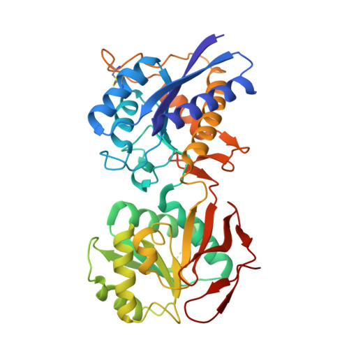
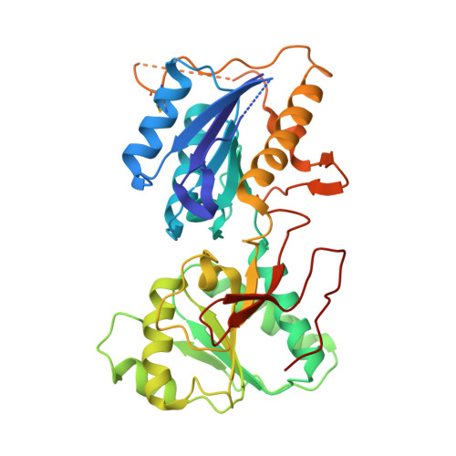
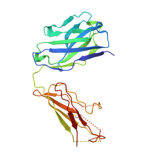
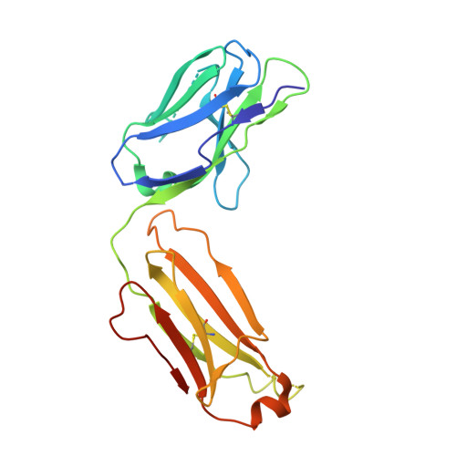
Molecular Basis for Subtype Specificity and High-Affinity Zinc Inhibition in the GluN1-GluN2A NMDA Receptor Amino-Terminal Domain.

Romero-Hernandez, A.Simorowski, N.Karakas, E.Furukawa, H.

(2016) Neuron 92: 1324-1336

  • DOI: https://doi.org/10.1016/j.neuron.2016.11.006
  • Primary Citation of Related Structures:  
    5TPW, 5TPZ, 5TQ0, 5TQ2

  • PubMed Abstract: 

    Zinc is vastly present in the mammalian brain and controls functions of various cell surface receptors to regulate neurotransmission. A distinctive characteristic of N-methyl-D-aspartate (NMDA) receptors containing a GluN2A subunit is that their ion channel activity is allosterically inhibited by a nano-molar concentration of zinc that binds to an extracellular domain called an amino-terminal domain (ATD). Despite physiological importance, the molecular mechanism underlying the high-affinity zinc inhibition has been incomplete because of the lack of a GluN2A ATD structure. Here we show the first crystal structures of the heterodimeric GluN1-GluN2A ATD, which provide the complete map of the high-affinity zinc-binding site and reveal distinctive features from the ATD of the GluN1-GluN2B subtype. Perturbation of hydrogen bond networks at the hinge of the GluN2A bi-lobe structure affects both zinc inhibition and open probability, supporting the general model in which the bi-lobe motion in ATD regulates the channel activity in NMDA receptors.


  • Organizational Affiliation

    WM Keck Structural Biology Laboratory, Cold Spring Harbor Laboratory, Cold Spring Harbor, NY 11724, USA; Watson School of Biological Sciences, Cold Spring Harbor, NY 11724, USA.


Macromolecules
Find similar proteins by:  (by identity cutoff)  |  3D Structure
Entity ID: 1
MoleculeChains Sequence LengthOrganismDetailsImage
NMDA glutamate receptor subunit389Xenopus laevisMutation(s): 2 
Gene Names: grin1NR1
UniProt
Find proteins for A0A1L8F5J9 (Xenopus laevis)
Explore A0A1L8F5J9 
Go to UniProtKB:  A0A1L8F5J9
Entity Groups  
Sequence Clusters30% Identity50% Identity70% Identity90% Identity95% Identity100% Identity
UniProt GroupA0A1L8F5J9
Glycosylation
Glycosylation Sites: 2
Sequence Annotations
Expand
  • Reference Sequence
Find similar proteins by:  (by identity cutoff)  |  3D Structure
Entity ID: 2
MoleculeChains Sequence LengthOrganismDetailsImage
Glutamate receptor ionotropic, NMDA 2A360Rattus norvegicusMutation(s): 0 
Gene Names: Grin2a
UniProt
Find proteins for Q00959 (Rattus norvegicus)
Explore Q00959 
Go to UniProtKB:  Q00959
Entity Groups  
Sequence Clusters30% Identity50% Identity70% Identity90% Identity95% Identity100% Identity
UniProt GroupQ00959
Sequence Annotations
Expand
  • Reference Sequence
Find similar proteins by:  (by identity cutoff)  |  3D Structure
Entity ID: 3
MoleculeChains Sequence LengthOrganismDetailsImage
FAB, HEAVY CHAINC [auth H]221Mus musculusMutation(s): 0 
Entity Groups  
Sequence Clusters30% Identity50% Identity70% Identity90% Identity95% Identity100% Identity
Sequence Annotations
Expand
  • Reference Sequence
Find similar proteins by:  (by identity cutoff)  |  3D Structure
Entity ID: 4
MoleculeChains Sequence LengthOrganismDetailsImage
FAB, LIGHT CHAIND [auth L]214Mus musculusMutation(s): 0 
Entity Groups  
Sequence Clusters30% Identity50% Identity70% Identity90% Identity95% Identity100% Identity
Sequence Annotations
Expand
  • Reference Sequence
Small Molecules
Ligands 4 Unique
IDChains Name / Formula / InChI Key2D Diagram3D Interactions
NAG
Query on NAG

Download Ideal Coordinates CCD File 
F [auth A],
G [auth A]
2-acetamido-2-deoxy-beta-D-glucopyranose
C8 H15 N O6
OVRNDRQMDRJTHS-FMDGEEDCSA-N
SO4
Query on SO4

Download Ideal Coordinates CCD File 
H [auth A],
I [auth A],
J [auth A],
M [auth B]
SULFATE ION
O4 S
QAOWNCQODCNURD-UHFFFAOYSA-L
GOL
Query on GOL

Download Ideal Coordinates CCD File 
K [auth A],
N [auth B]
GLYCEROL
C3 H8 O3
PEDCQBHIVMGVHV-UHFFFAOYSA-N
ZN
Query on ZN

Download Ideal Coordinates CCD File 
E [auth A],
L [auth B]
ZINC ION
Zn
PTFCDOFLOPIGGS-UHFFFAOYSA-N
Experimental Data & Validation

Experimental Data

  • Method: X-RAY DIFFRACTION
  • Resolution: 3.29 Å
  • R-Value Free: 0.261 
  • R-Value Work: 0.224 
  • R-Value Observed: 0.228 
  • Space Group: P 21 21 21
Unit Cell:
Length ( Å )Angle ( ˚ )
a = 78.163α = 90
b = 117.826β = 90
c = 158.992γ = 90
Software Package:
Software NamePurpose
PHENIXrefinement
HKL-2000data collection
PDB_EXTRACTdata extraction
HKL-2000data reduction
PHASERphasing
DENZOdata reduction
SCALEPACKdata scaling

Structure Validation

View Full Validation Report


Ligand Structure Quality Assessment 


Entry History & Funding Information

Deposition Data


Funding OrganizationLocationGrant Number
National Institutes of Health/National Institute of Mental Health (NIH/NIMH)United StatesMH085926
National Institutes of Health/National Institute of General Medical Sciences (NIH/NIGMS)United StatesGM105730

Revision History  (Full details and data files)

  • Version 1.0: 2016-12-14
    Type: Initial release
  • Version 1.1: 2017-09-20
    Changes: Author supporting evidence, Refinement description
  • Version 1.2: 2018-05-16
    Changes: Data collection, Database references
  • Version 1.3: 2019-11-27
    Changes: Author supporting evidence
  • Version 1.4: 2020-07-29
    Type: Remediation
    Reason: Carbohydrate remediation
    Changes: Data collection, Derived calculations, Structure summary
  • Version 1.5: 2023-10-04
    Changes: Data collection, Database references, Refinement description, Structure summary