5TPR | pdb_00005tpr

Desmethyl-4-deoxygadusol synthase from Anabaena variabilis (Ava_3858) with NAD+ and Zn2+ bound


Experimental Data Snapshot

  • Method: X-RAY DIFFRACTION
  • Resolution: 1.70 Å
  • R-Value Free: 
    0.183 (Depositor), 0.160 (DCC) 
  • R-Value Work: 
    0.156 (Depositor) 
  • R-Value Observed: 
    0.157 (Depositor) 

Starting Model: experimental
View more details

wwPDB Validation 3D Report Full Report

Validation slider image for 5TPR

Ligand Structure Quality Assessment 


This is version 1.2 of the entry. See complete history

Literature

Evolution and Distribution of C7-Cyclitol Synthases in Prokaryotes and Eukaryotes.

Osborn, A.R.Kean, K.M.Alseud, K.M.Almabruk, K.H.Asamizu, S.Lee, J.A.Karplus, P.A.Mahmud, T.

(2017) ACS Chem Biol 12: 979-988

  • DOI: https://doi.org/10.1021/acschembio.7b00066
  • Primary Citation Related Structures: 
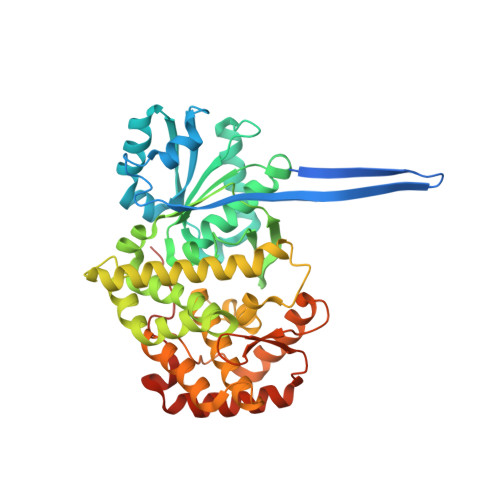
    5TPR

  • PubMed Abstract: 

    2-Epi-5-epi-valiolone synthase (EEVS), a C 7 -sugar phosphate cyclase (SPC) homologous to 3-dehydroquinate synthase (DHQS), was discovered during studies of the biosynthesis of the C 7 N-aminocyclitol family of natural products. EEVS was originally thought to be present only in certain actinomycetes, but analyses of genome sequences showed that it is broadly distributed in both prokaryotes and eukaryotes, including vertebrates. Another SPC, desmethyl-4-deoxygadusol synthase (DDGS), was later discovered as being involved in the biosynthesis of mycosporine-like amino acid sunscreen compounds. Current database annotations are quite unreliable, with many EEVSs reported as DHQS, and most DDGSs reported as EEVS, DHQS, or simply hypothetical proteins. Here, we identify sequence features useful for distinguishing these enzymes, report a crystal structure of a representative DDGS showing the high similarity of the EEVS and DDGS enzymes, identify notable active site differences, and demonstrate the importance of two of these active site residues for catalysis by point mutations. Further, we functionally characterized two representatives of a distinct clade equidistant from known EEVS and known DDGS groups and show them to be authentic EEVSs. Moreover, we document and discuss the distribution of genes that encode EEVS and DDGS in various prokaryotes and eukaryotes, including pathogenic bacteria, plant symbionts, nitrogen-fixing bacteria, myxobacteria, cyanobacteria, fungi, stramenopiles, and animals, suggesting their broad potential biological roles in nature.


  • Organizational Affiliation
    • Department of Pharmaceutical Sciences, Oregon State University , Corvallis, Oregon 97331-3507, United States.

Macromolecule Content 

  • Total Structure Weight: 102.7 kDa 
  • Atom Count: 7,961 
  • Modeled Residue Count: 800 
  • Deposited Residue Count: 888 
  • Unique protein chains: 1

Macromolecules

Find similar proteins by:|  3D Structure
Entity ID: 1
MoleculeChains  Sequence LengthOrganismDetailsImage
3-dehydroquinate synthase
A, B
444Trichormus variabilis ATCC 29413Mutation(s): 0 
Gene Names: Ava_3858
EC: 4.2.3.4 (PDB Primary Data), 4.2.3.154 (UniProt)
UniProt
Find proteins for Q3M6C3 (Trichormus variabilis (strain ATCC 29413 / PCC 7937))
Explore Q3M6C3 
Go to UniProtKB:  Q3M6C3
Entity Groups
Sequence Clusters30% Identity50% Identity70% Identity90% Identity95% Identity100% Identity
UniProt GroupQ3M6C3
Sequence Annotations
Expand
Reference Sequence

Small Molecules

Ligands 5 Unique
IDChains Name / Formula / InChI Key2D Diagram3D Interactions
NAD

Query on NAD


Download:Ideal Coordinates CCD File
E [auth A],
K [auth B]
NICOTINAMIDE-ADENINE-DINUCLEOTIDE
C21 H27 N7 O14 P2
BAWFJGJZGIEFAR-NNYOXOHSSA-N
PEG

Query on PEG


Download:Ideal Coordinates CCD File
H [auth A],
N [auth B]
DI(HYDROXYETHYL)ETHER
C4 H10 O3
MTHSVFCYNBDYFN-UHFFFAOYSA-N
SO4

Query on SO4


Download:Ideal Coordinates CCD File
D [auth A]
F [auth A]
G [auth A]
L [auth B]
M [auth B]
D [auth A],
F [auth A],
G [auth A],
L [auth B],
M [auth B],
P [auth B]
SULFATE ION
O4 S
QAOWNCQODCNURD-UHFFFAOYSA-L
GOL

Query on GOL


Download:Ideal Coordinates CCD File
I [auth A],
O [auth B]
GLYCEROL
C3 H8 O3
PEDCQBHIVMGVHV-UHFFFAOYSA-N
ZN

Query on ZN


Download:Ideal Coordinates CCD File
C [auth A],
J [auth B]
ZINC ION
Zn
PTFCDOFLOPIGGS-UHFFFAOYSA-N

Experimental Data & Validation

Experimental Data

  • Method: X-RAY DIFFRACTION
  • Resolution: 1.70 Å
  • R-Value Free:  0.183 (Depositor), 0.160 (DCC) 
  • R-Value Work:  0.156 (Depositor) 
  • R-Value Observed: 0.157 (Depositor) 
Space Group: P 21 21 21
Unit Cell:
Length ( Å )Angle ( ˚ )
a = 66.5α = 90
b = 120.261β = 90
c = 133.26γ = 90
Software Package:
Software NamePurpose
PHENIXrefinement
Aimlessdata scaling
MR-Rosettaphasing
iMOSFLMdata reduction

Structure Validation

View Full Validation Report


Ligand Structure Quality Assessment 


Entry History 

Deposition Data

Revision History  (Full details and data files)

  • Version 1.0: 2017-02-22
    Type: Initial release
  • Version 1.1: 2017-05-03
    Changes: Data collection, Database references
  • Version 1.2: 2023-10-04
    Changes: Data collection, Database references, Derived calculations, Refinement description