5TKB | pdb_00005tkb

CRYSTAL STRUCTURE OF HUMAN PHOSPHODIESTERASE 4D IN COMPLEX WITH A TETRAFLUORANLINE COMPOUND


Experimental Data Snapshot

  • Method: X-RAY DIFFRACTION
  • Resolution: 2.16 Å
  • R-Value Free: 
    0.209 (Depositor), 0.211 (DCC) 
  • R-Value Work: 
    0.188 (Depositor), 0.190 (DCC) 
  • R-Value Observed: 
    0.189 (Depositor) 

Starting Model: experimental
View more details

wwPDB Validation 3D Report Full Report

Validation slider image for 5TKB

Ligand Structure Quality Assessment 


This is version 1.2 of the entry. See complete history

Literature

Identification of imidazo[1,2-b]pyridazine TYK2 pseudokinase ligands as potent and selective allosteric inhibitors of TYK2 signalling.

Moslin, R.Gardner, D.Santella, J.Zhang, Y.Duncia, J.V.Liu, C.Lin, J.Tokarski, J.S.Strnad, J.Pedicord, D.Chen, J.Blat, Y.Zupa-Fernandez, A.Cheng, L.Sun, H.Chaudhry, C.Huang, C.D'Arienzo, C.Sack, J.S.Muckelbauer, J.K.Chang, C.Tredup, J.Xie, D.Aranibar, N.Burke, J.R.Carter, P.H.Weinstein, D.S.

(2017) Medchemcomm 8: 700-712

  • DOI: https://doi.org/10.1039/c6md00560h
  • Primary Citation Related Structures: 
    5TKB, 5TKD

  • PubMed Abstract: 

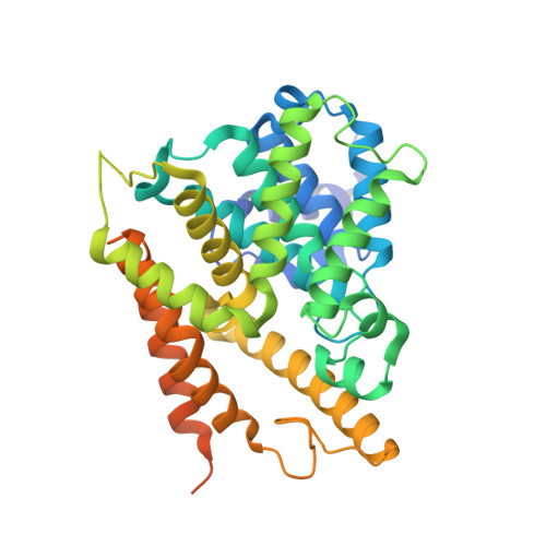
    As a member of the Janus (JAK) family of non-receptor tyrosine kinases, TYK2 mediates the signaling of pro-inflammatory cytokines including IL-12, IL-23 and type 1 interferon (IFN), and therefore represents an attractive potential target for treating the various immuno-inflammatory diseases in which these cytokines have been shown to play a role. Following up on our previous report that ligands to the pseudokinase domain (JH2) of TYK2 suppress cytokine-mediated receptor activation of the catalytic (JH1) domain, the imidazo[1,2- b ]pyridazine (IZP) 7 was identified as a promising hit compound. Through iterative modification of each of the substituents of the IZP scaffold, the cellular potency was improved while maintaining selectivity over the JH1 domain. These studies led to the discovery of the JH2-selective TYK2 inhibitor 29 , which provided encouraging systemic exposures after oral dosing in mice. Phosphodiesterase 4 (PDE4) was identified as an off-target and potential liability of the IZP ligands, and selectivity for TYK2 JH2 over this enzyme was obtained by elaborating along selectivity vectors determined from analyses of X-ray co-crystal structures of representative ligands of the IZP class bound to both proteins.


  • Organizational Affiliation
    • Bristol-Myers Squibb Research , Princeton , New Jersey , USA . Email: ryan.moslin@bms.com.

Macromolecule Content 

  • Total Structure Weight: 177.95 kDa 
  • Atom Count: 11,083 
  • Modeled Residue Count: 1,297 
  • Deposited Residue Count: 1,524 
  • Unique protein chains: 1

Macromolecules

Find similar proteins by:|  3D Structure
Entity ID: 1
MoleculeChains  Sequence LengthOrganismDetailsImage
cAMP-specific 3',5'-cyclic phosphodiesterase 4D
A, B, C, D
381Homo sapiensMutation(s): 2 
Gene Names: PDE4DDPDE3
EC: 3.1.4.53
UniProt & NIH Common Fund Data Resources
Find proteins for Q08499 (Homo sapiens)
Explore Q08499 
Go to UniProtKB:  Q08499
PHAROS:  Q08499
GTEx:  ENSG00000113448 
Entity Groups
Sequence Clusters30% Identity50% Identity70% Identity90% Identity95% Identity100% Identity
UniProt GroupQ08499
Sequence Annotations
Expand
Reference Sequence

Small Molecules

Ligands 5 Unique
IDChains Name / Formula / InChI Key2D Diagram3D Interactions
7DJ

Query on 7DJ


Download:Ideal Coordinates CCD File
AA [auth D],
K [auth A],
R [auth B],
W [auth C]
N-[(2R)-2,3-dihydroxy-2-methylpropyl]-8-(methylamino)-6-[(2,3,5,6-tetrafluorophenyl)amino]imidazo[1,2-b]pyridazine-3-carboxamide
C18 H18 F4 N6 O3
HNSFAKGAIZARCR-GOSISDBHSA-N
SO4

Query on SO4


Download:Ideal Coordinates CCD File
G [auth A],
N [auth B],
U [auth C]
SULFATE ION
O4 S
QAOWNCQODCNURD-UHFFFAOYSA-L
ZN

Query on ZN


Download:Ideal Coordinates CCD File
E [auth A],
L [auth B],
S [auth C],
X [auth D]
ZINC ION
Zn
PTFCDOFLOPIGGS-UHFFFAOYSA-N
EOH

Query on EOH


Download:Ideal Coordinates CCD File
H [auth A]
I [auth A]
J [auth A]
O [auth B]
P [auth B]
H [auth A],
I [auth A],
J [auth A],
O [auth B],
P [auth B],
Q [auth B],
V [auth C],
Z [auth D]
ETHANOL
C2 H6 O
LFQSCWFLJHTTHZ-UHFFFAOYSA-N
MG

Query on MG


Download:Ideal Coordinates CCD File
F [auth A],
M [auth B],
T [auth C],
Y [auth D]
MAGNESIUM ION
Mg
JLVVSXFLKOJNIY-UHFFFAOYSA-N

Experimental Data & Validation

Experimental Data

  • Method: X-RAY DIFFRACTION
  • Resolution: 2.16 Å
  • R-Value Free:  0.209 (Depositor), 0.211 (DCC) 
  • R-Value Work:  0.188 (Depositor), 0.190 (DCC) 
  • R-Value Observed: 0.189 (Depositor) 
Space Group: P 21 21 21
Unit Cell:
Length ( Å )Angle ( ˚ )
a = 99α = 90
b = 112.7β = 90
c = 159.88γ = 90
Software Package:
Software NamePurpose
XDSdata reduction
Aimlessdata scaling
BUSTERrefinement
BUSTERphasing

Structure Validation

View Full Validation Report


Ligand Structure Quality Assessment 


Entry History 

Deposition Data

  • Released Date: 2016-12-28 
  • Deposition Author(s): Sack, J.S.

Revision History  (Full details and data files)

  • Version 1.0: 2016-12-28
    Type: Initial release
  • Version 1.1: 2018-08-29
    Changes: Data collection, Database references
  • Version 1.2: 2023-10-04
    Changes: Data collection, Database references, Derived calculations, Refinement description