5TJZ | pdb_00005tjz

Structure of 4-Hydroxytetrahydrodipicolinate Reductase from Mycobacterium tuberculosis with NADPH and 2,6 Pyridine Dicarboxylic Acid


Experimental Data Snapshot

  • Method: X-RAY DIFFRACTION
  • Resolution: 1.50 Å
  • R-Value Free: 
    0.169 (Depositor), 0.171 (DCC) 
  • R-Value Work: 
    0.148 (Depositor), 0.151 (DCC) 
  • R-Value Observed: 
    0.149 (Depositor) 

Starting Model: experimental
View more details

wwPDB Validation 3D Report Full Report

Validation slider image for 5TJZ

Ligand Structure Quality Assessment 


This is version 1.2 of the entry. See complete history

Literature

Comparative structural and mechanistic studies of 4-hydroxy-tetrahydrodipicolinate reductases from Mycobacterium tuberculosis and Vibrio vulnificus.

Pote, S.Kachhap, S.Mank, N.J.Daneshian, L.Klapper, V.Pye, S.Arnette, A.K.Shimizu, L.S.Borowski, T.Chruszcz, M.

(2021) Biochim Biophys Acta Gen Subj 1865: 129750-129750

  • DOI: https://doi.org/10.1016/j.bbagen.2020.129750
  • Primary Citation Related Structures: 
    5TEJ, 5TEK, 5TEM, 5TEN, 5TJY, 5TJZ, 5UGV, 5US6

  • PubMed Abstract: 

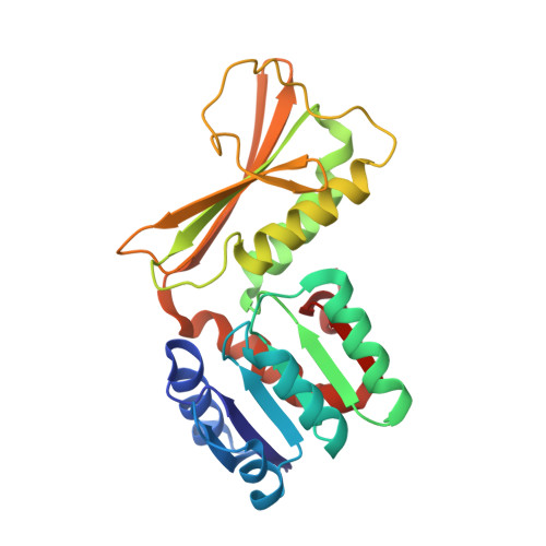
    The products of the lysine biosynthesis pathway, meso-diaminopimelate and lysine, are essential for bacterial survival. This paper focuses on the structural and mechanistic characterization of 4-hydroxy-tetrahydrodipicolinate reductase (DapB), which is one of the enzymes from the lysine biosynthesis pathway. DapB catalyzes the conversion of (2S, 4S)-4-hydroxy-2,3,4,5-tetrahydrodipicolinate (HTPA) to 2,3,4,5-tetrahydrodipicolinate in an NADH/NADPH dependent reaction. Genes coding for DapBs were identified as essential for many pathogenic bacteria, and therefore DapB is an interesting new target for the development of antibiotics. We have combined experimental and computational approaches to provide novel insights into mechanism of the DapB catalyzed reaction. Structures of DapBs originating from Mycobacterium tuberculosis and Vibrio vulnificus in complexes with NAD + , NADP + , as well as with inhibitors, were determined and described. The structures determined by us, as well as currently available structures of DapBs from other bacterial species, were compared and used to elucidate a mechanism of reaction catalyzed by this group of enzymes. Several different computational methods were used to provide a detailed description of a plausible reaction mechanism. This is the first report presenting the detailed mechanism of reaction catalyzed by DapB. Structural data in combination with information on the reaction mechanism provide a background for development of DapB inhibitors, including transition-state analogues.


  • Organizational Affiliation
    • Department of Chemistry and Biochemistry, University of South Carolina, Columbia, SC 29208, USA.

Macromolecule Content 

  • Total Structure Weight: 27.68 kDa 
  • Atom Count: 2,331 
  • Modeled Residue Count: 245 
  • Deposited Residue Count: 245 
  • Unique protein chains: 1

Macromolecules

Find similar proteins by:|  3D Structure
Entity ID: 1
MoleculeChains  Sequence LengthOrganismDetailsImage
4-hydroxy-tetrahydrodipicolinate reductase245Mycobacterium tuberculosis H37RaMutation(s): 0 
Gene Names: dapBMRA_2798
EC: 1.17.1.8
UniProt
Find proteins for P9WP23 (Mycobacterium tuberculosis (strain ATCC 25618 / H37Rv))
Explore P9WP23 
Go to UniProtKB:  P9WP23
Entity Groups
Sequence Clusters30% Identity50% Identity70% Identity90% Identity95% Identity100% Identity
UniProt GroupP9WP23
Sequence Annotations
Expand
Reference Sequence

Small Molecules

Ligands 8 Unique
IDChains Name / Formula / InChI Key2D Diagram3D Interactions
NAP

Query on NAP


Download:Ideal Coordinates CCD File
E [auth A]NADP NICOTINAMIDE-ADENINE-DINUCLEOTIDE PHOSPHATE
C21 H28 N7 O17 P3
XJLXINKUBYWONI-NNYOXOHSSA-N
PDC

Query on PDC


Download:Ideal Coordinates CCD File
B [auth A]PYRIDINE-2,6-DICARBOXYLIC ACID
C7 H5 N O4
WJJMNDUMQPNECX-UHFFFAOYSA-N
AE3

Query on AE3


Download:Ideal Coordinates CCD File
F [auth A]2-(2-ETHOXYETHOXY)ETHANOL
C6 H14 O3
XXJWXESWEXIICW-UHFFFAOYSA-N
BEZ

Query on BEZ


Download:Ideal Coordinates CCD File
C [auth A]BENZOIC ACID
C7 H6 O2
WPYMKLBDIGXBTP-UHFFFAOYSA-N
SO4

Query on SO4


Download:Ideal Coordinates CCD File
P [auth A]SULFATE ION
O4 S
QAOWNCQODCNURD-UHFFFAOYSA-L
IMD

Query on IMD


Download:Ideal Coordinates CCD File
D [auth A]IMIDAZOLE
C3 H5 N2
RAXXELZNTBOGNW-UHFFFAOYSA-O
EDO

Query on EDO


Download:Ideal Coordinates CCD File
H [auth A]
I [auth A]
J [auth A]
K [auth A]
L [auth A]
H [auth A],
I [auth A],
J [auth A],
K [auth A],
L [auth A],
M [auth A],
N [auth A],
O [auth A],
S [auth A]
1,2-ETHANEDIOL
C2 H6 O2
LYCAIKOWRPUZTN-UHFFFAOYSA-N
CL

Query on CL


Download:Ideal Coordinates CCD File
G [auth A],
Q [auth A],
R [auth A]
CHLORIDE ION
Cl
VEXZGXHMUGYJMC-UHFFFAOYSA-M

Experimental Data & Validation

Experimental Data

  • Method: X-RAY DIFFRACTION
  • Resolution: 1.50 Å
  • R-Value Free:  0.169 (Depositor), 0.171 (DCC) 
  • R-Value Work:  0.148 (Depositor), 0.151 (DCC) 
  • R-Value Observed: 0.149 (Depositor) 
Space Group: P 62 2 2
Unit Cell:
Length ( Å )Angle ( ˚ )
a = 67.157α = 90
b = 67.157β = 90
c = 251.325γ = 120
Software Package:
Software NamePurpose
REFMACrefinement
HKL-3000data collection
HKL-2000data scaling
PDB_EXTRACTdata extraction
HKL-3000data reduction
HKL-3000phasing

Structure Validation

View Full Validation Report


Ligand Structure Quality Assessment 


Entry History 

Deposition Data

Revision History  (Full details and data files)

  • Version 1.0: 2018-01-10
    Type: Initial release
  • Version 1.1: 2021-06-30
    Changes: Database references
  • Version 1.2: 2023-10-04
    Changes: Data collection, Database references, Refinement description