5TIH | pdb_00005tih

Structural basis for inhibition of erythrocyte invasion by antibodies to Plasmodium falciparum protein CyRPA


Experimental Data Snapshot

  • Method: X-RAY DIFFRACTION
  • Resolution: 2.44 Å
  • R-Value Free: 
    0.211 (Depositor), 0.213 (DCC) 
  • R-Value Work: 
    0.189 (Depositor), 0.193 (DCC) 
  • R-Value Observed: 
    0.190 (Depositor) 

Starting Model: experimental
View more details

wwPDB Validation 3D Report Full Report

Validation slider image for 5TIH

This is version 1.6 of the entry. See complete history

Literature

Structural basis for inhibition of erythrocyte invasion by antibodies toPlasmodium falciparumprotein CyRPA.

Chen, L.Xu, Y.Wong, W.Thompson, J.K.Healer, J.Goddard-Borger, E.D.Lawrence, M.C.Cowman, A.F.

(2017) Elife 6

  • DOI: https://doi.org/10.7554/eLife.21347
  • Primary Citation Related Structures: 
    5TIH, 5TIK

  • PubMed Abstract: 

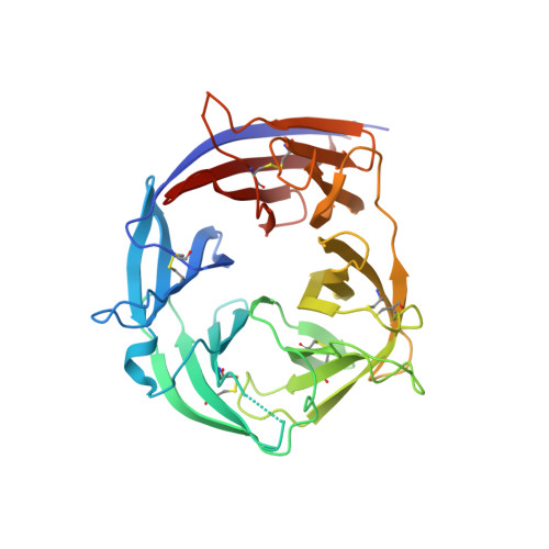
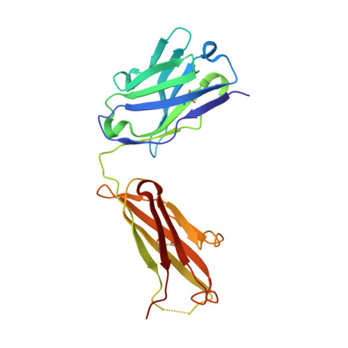
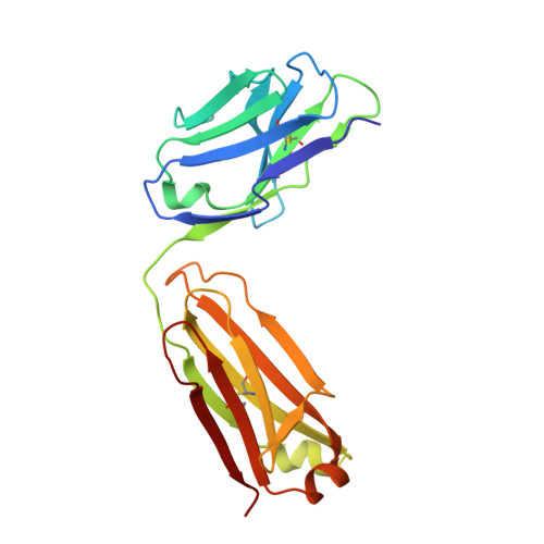
    Plasmodium falciparum causes malaria in humans with over 450,000 deaths annually. The asexual blood stage involves invasion of erythrocytes by merozoites, in which they grow and divide to release daughter merozoites, which in turn invade new erythrocytes perpetuating the cycle responsible for malaria. A key step in merozoite invasion is the essential binding of PfRh5/CyRPA/PfRipr complex to basigin, a step linked to the formation of a pore between merozoites and erythrocytes. We show CyRPA interacts directly with PfRh5. An invasion inhibitory monoclonal antibody to CyRPA blocks binding of CyRPA to PfRh5 and complex formation thus illuminating the molecular mechanism for inhibition of parasite growth. We determined the crystal structures of CyRPA alone and in complex with an antibody Fab fragment. CyRPA has a six-bladed β-propeller fold, and we identify the region that interacts with PfRh5. This functionally conserved epitope is a potential target for vaccines against P. falciparum .


  • Organizational Affiliation
    • The Walter and Eliza Hall Institute of Medical Research, Melbourne, Australia.

Macromolecule Content 

  • Total Structure Weight: 86.54 kDa 
  • Atom Count: 6,045 
  • Modeled Residue Count: 750 
  • Deposited Residue Count: 762 
  • Unique protein chains: 3

Macromolecules

Find similar proteins by:|  3D Structure
Entity ID: 1
MoleculeChains  Sequence LengthOrganismDetailsImage
Cysteine-rich protective antigen335Plasmodium falciparum 3D7Mutation(s): 0 
Gene Names: PF3D7_0423800
UniProt
Find proteins for Q8IFM8 (Plasmodium falciparum (isolate 3D7))
Explore Q8IFM8 
Go to UniProtKB:  Q8IFM8
Entity Groups
Sequence Clusters30% Identity50% Identity70% Identity90% Identity95% Identity100% Identity
UniProt GroupQ8IFM8
Sequence Annotations
Expand
Reference Sequence
Find similar proteins by:|  3D Structure
Entity ID: 2
MoleculeChains  Sequence LengthOrganismDetailsImage
CyRPA antibody Fab Heavy ChainB [auth H]215Mus musculusMutation(s): 0 
Find similar proteins by:|  3D Structure
Entity ID: 3
MoleculeChains  Sequence LengthOrganismDetailsImage
CyRPA antibody Fab Light ChainC [auth L]212Mus musculusMutation(s): 0 

Experimental Data & Validation

Experimental Data

  • Method: X-RAY DIFFRACTION
  • Resolution: 2.44 Å
  • R-Value Free:  0.211 (Depositor), 0.213 (DCC) 
  • R-Value Work:  0.189 (Depositor), 0.193 (DCC) 
  • R-Value Observed: 0.190 (Depositor) 
Space Group: P 21 21 21
Unit Cell:
Length ( Å )Angle ( ˚ )
a = 79.95α = 90
b = 87.38β = 90
c = 145.14γ = 90
Software Package:
Software NamePurpose
PHENIXrefinement
XSCALEdata scaling
PDB_EXTRACTdata extraction
XDSdata reduction
PHASERphasing

Structure Validation

View Full Validation Report


Entry History 

& Funding Information

Deposition Data


Funding OrganizationLocationGrant Number
National Health and Medical Research Council (NHMRC, Australia)Australia--

Revision History  (Full details and data files)

  • Version 1.0: 2017-03-01
    Type: Initial release
  • Version 1.1: 2017-09-13
    Changes: Author supporting evidence, Data collection
  • Version 1.2: 2018-02-21
    Changes: Database references
  • Version 1.3: 2018-03-28
    Changes: Data collection, Database references
  • Version 1.4: 2020-01-08
    Changes: Author supporting evidence
  • Version 1.5: 2023-10-04
    Changes: Data collection, Database references, Refinement description
  • Version 1.6: 2024-10-23
    Changes: Structure summary