5TIA

Crystal structure of human TDO in complex with Trp, Northeast Structural Genomics Consortium Target HR6161


Experimental Data Snapshot

  • Method: X-RAY DIFFRACTION
  • Resolution: 2.44 Å
  • R-Value Free: 0.230 
  • R-Value Work: 0.168 
  • R-Value Observed: 0.174 

Starting Model: experimental
View more details

wwPDB Validation   3D Report Full Report


Ligand Structure Quality Assessment 


This is version 1.7 of the entry. See complete history


Literature

Molecular basis for catalysis and substrate-mediated cellular stabilization of human tryptophan 2,3-dioxygenase.

Lewis-Ballester, A.Forouhar, F.Kim, S.M.Lew, S.Wang, Y.Karkashon, S.Seetharaman, J.Batabyal, D.Chiang, B.Y.Hussain, M.Correia, M.A.Yeh, S.R.Tong, L.

(2016) Sci Rep 6: 35169-35169

  • DOI: https://doi.org/10.1038/srep35169
  • Primary Citation of Related Structures:  
    5TI9, 5TIA

  • PubMed Abstract: 

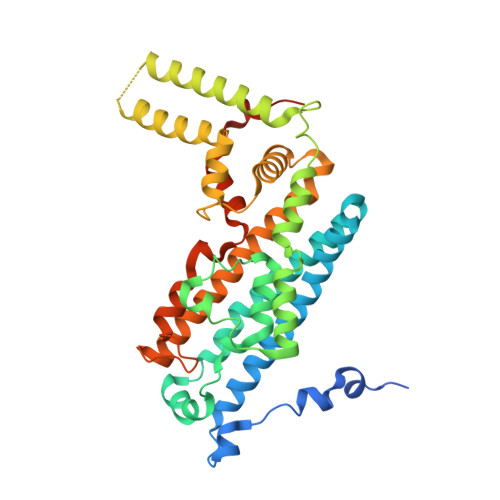
    Tryptophan 2,3-dioxygenase (TDO) and indoleamine 2,3-dioxygenase (IDO) play a central role in tryptophan metabolism and are involved in many cellular and disease processes. Here we report the crystal structure of human TDO (hTDO) in a ternary complex with the substrates L-Trp and O 2 and in a binary complex with the product N-formylkynurenine (NFK), defining for the first time the binding modes of both substrates and the product of this enzyme. The structure indicates that the dioxygenation reaction is initiated by a direct attack of O 2 on the C 2 atom of the L-Trp indole ring. The structure also reveals an exo binding site for L-Trp, located ~42 Å from the active site and formed by residues conserved among tryptophan-auxotrophic TDOs. Biochemical and cellular studies indicate that Trp binding at this exo site does not affect enzyme catalysis but instead it retards the degradation of hTDO through the ubiquitin-dependent proteasomal pathway. This exo site may therefore provide a novel L-Trp-mediated regulation mechanism for cellular degradation of hTDO, which may have important implications in human diseases.


  • Organizational Affiliation

    Department of Physiology and Biophysics Albert Einstein College of Medicine Bronx, NY 10461, USA.


Macromolecules
Find similar proteins by:  (by identity cutoff)  |  3D Structure
Entity ID: 1
MoleculeChains Sequence LengthOrganismDetailsImage
Tryptophan 2,3-dioxygenase
A, B, C, D
380Homo sapiensMutation(s): 0 
Gene Names: TDO2TDO
EC: 1.13.11.11
UniProt & NIH Common Fund Data Resources
Find proteins for P48775 (Homo sapiens)
Explore P48775 
Go to UniProtKB:  P48775
GTEx:  ENSG00000151790 
Entity Groups  
Sequence Clusters30% Identity50% Identity70% Identity90% Identity95% Identity100% Identity
UniProt GroupP48775
Sequence Annotations
Expand
  • Reference Sequence
Small Molecules
Ligands 2 Unique
IDChains Name / Formula / InChI Key2D Diagram3D Interactions
HEM
Query on HEM

Download Ideal Coordinates CCD File 
E [auth A],
H [auth B],
K [auth C],
N [auth D]
PROTOPORPHYRIN IX CONTAINING FE
C34 H32 Fe N4 O4
KABFMIBPWCXCRK-RGGAHWMASA-L
TRP
Query on TRP

Download Ideal Coordinates CCD File 
F [auth A]
G [auth A]
I [auth B]
J [auth B]
L [auth C]
F [auth A],
G [auth A],
I [auth B],
J [auth B],
L [auth C],
M [auth C],
O [auth D],
P [auth D]
TRYPTOPHAN
C11 H12 N2 O2
QIVBCDIJIAJPQS-VIFPVBQESA-N
Experimental Data & Validation

Experimental Data

  • Method: X-RAY DIFFRACTION
  • Resolution: 2.44 Å
  • R-Value Free: 0.230 
  • R-Value Work: 0.168 
  • R-Value Observed: 0.174 
  • Space Group: P 21 21 2
Unit Cell:
Length ( Å )Angle ( ˚ )
a = 144.359α = 90
b = 153.561β = 90
c = 88.163γ = 90
Software Package:
Software NamePurpose
PHENIXrefinement
HKL-2000data reduction
HKL-2000data scaling
MOLREPphasing

Structure Validation

View Full Validation Report


Ligand Structure Quality Assessment 


Entry History & Funding Information

Deposition Data


Funding OrganizationLocationGrant Number
National Institutes of Health/National Institute of General Medical Sciences (NIH/NIGMS)United StatesP50 GM62413
National Institutes of Health/National Institute of General Medical Sciences (NIH/NIGMS)United StatesU54 GM074958

Revision History  (Full details and data files)

  • Version 1.0: 2016-10-26
    Type: Initial release
  • Version 1.1: 2016-11-02
    Changes: Database references
  • Version 1.2: 2016-11-09
    Changes: Structure summary
  • Version 1.3: 2016-11-30
    Changes: Structure summary
  • Version 1.4: 2016-12-14
    Changes: Structure summary
  • Version 1.5: 2017-09-27
    Changes: Author supporting evidence
  • Version 1.6: 2019-12-25
    Changes: Author supporting evidence
  • Version 1.7: 2023-10-04
    Changes: Data collection, Database references, Refinement description