5THO | pdb_00005tho

Crystal Structure of Mycobacterium Tuberculosis Proteasome in complex with N,C-capped Dipeptide Inhibitor PKS2205


Experimental Data Snapshot

  • Method: X-RAY DIFFRACTION
  • Resolution: 3.00 Å
  • R-Value Free: 
    0.210 (Depositor), 0.210 (DCC) 
  • R-Value Work: 
    0.176 (Depositor), 0.180 (DCC) 
  • R-Value Observed: 
    0.178 (Depositor) 

Starting Model: experimental
View more details

wwPDB Validation 3D Report Full Report

Validation slider image for 5THO

Ligand Structure Quality Assessment 


This is version 1.3 of the entry. See complete history

Literature

Structural Basis for the Species-Selective Binding of N,C-Capped Dipeptides to the Mycobacterium tuberculosis Proteasome.

Hsu, H.C.Singh, P.K.Fan, H.Wang, R.Sukenick, G.Nathan, C.Lin, G.Li, H.

(2017) Biochemistry 56: 324-333

  • DOI: https://doi.org/10.1021/acs.biochem.6b01107
  • Primary Citation Related Structures: 
    5THO, 5TRG, 5TRR, 5TRS, 5TRY, 5TS0

  • PubMed Abstract: 

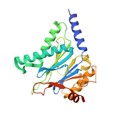
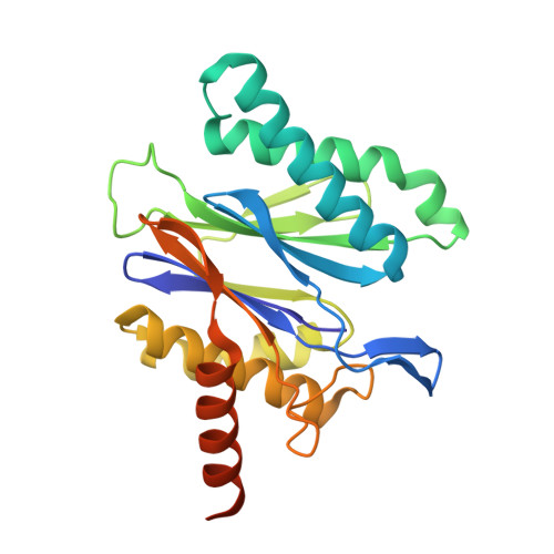
    The Mycobacterium tuberculosis (Mtb) 20S proteasome is vital for the pathogen to survive under nitrosative stress in vitro and to persist in mice. To qualify for drug development, inhibitors targeting Mtb 20S must spare both the human constitutive proteasome (c-20S) and immunoproteasome (i-20S). We recently reported members of a family of noncovalently binding dipeptide proteasome inhibitors that are highly potent and selective for Mtb 20S over human c-20S and i-20S. To understand the structural basis of their potency and selectivity, we have studied the structure-activity relationship of six derivatives and solved their cocrystal structures with Mtb 20S. The dipeptide inhibitors form an antiparallel β-strand with the active site β-strands. Selectivity is conferred by several features of Mtb 20S relative to its mouse counterparts, including a larger S1 pocket, additional hydrogen bonds in the S3 pocket, and hydrophobic interactions in the S4 pocket. Serine-20 and glutamine-22 of Mtb 20S interact with the dipeptides and confer Mtb-specific inhibition over c-20S and i-20S. The Mtb 20S and mammalian i-20S have a serine-27 that interacts strongly with the dipeptides, potentially explaining the higher inhibitory activity of the dipeptides toward i-20S over c-20S. This detailed structural knowledge will aid in optimizing the dipeptides as anti-tuberculosis drugs.


  • Organizational Affiliation
    • Van Andel Research Institute , Grand Rapids, Michigan 49503, United States.

Macromolecule Content 

  • Total Structure Weight: 725.3 kDa 
  • Atom Count: 47,201 
  • Modeled Residue Count: 6,157 
  • Deposited Residue Count: 6,720 
  • Unique protein chains: 2

Macromolecules

Find similar proteins by:|  3D Structure
Entity ID: 1
MoleculeChains  Sequence LengthOrganismDetailsImage
Proteasome subunit alpha
A, B, C, D, E
A, B, C, D, E, F, G, O, P, Q, R, S, T, U
240Mycobacterium tuberculosis H37RaMutation(s): 0 
Gene Names: prcAMRA_2124
EC: 3.4.25.1
UniProt
Find proteins for A5U4D5 (Mycobacterium tuberculosis (strain ATCC 25177 / H37Ra))
Explore A5U4D5 
Go to UniProtKB:  A5U4D5
Entity Groups
Sequence Clusters30% Identity50% Identity70% Identity90% Identity95% Identity100% Identity
UniProt GroupA5U4D5
Sequence Annotations
Expand
Reference Sequence
Find similar proteins by:|  3D Structure
Entity ID: 2
MoleculeChains  Sequence LengthOrganismDetailsImage
Proteasome subunit beta240Mycobacterium tuberculosis H37RaMutation(s): 0 
Gene Names: prcBMRA_2125
EC: 3.4.25.1
UniProt
Find proteins for A5U4D6 (Mycobacterium tuberculosis (strain ATCC 25177 / H37Ra))
Explore A5U4D6 
Go to UniProtKB:  A5U4D6
Entity Groups
Sequence Clusters30% Identity50% Identity70% Identity90% Identity95% Identity100% Identity
UniProt GroupA5U4D6
Sequence Annotations
Expand
Reference Sequence

Small Molecules

Ligands 1 Unique
IDChains Name / Formula / InChI Key2D Diagram3D Interactions
7C7

Query on 7C7


Download:Ideal Coordinates CCD File
CA [auth H]
DA [auth I]
EA [auth J]
FA [auth K]
GA [auth L]
CA [auth H],
DA [auth I],
EA [auth J],
FA [auth K],
GA [auth L],
HA [auth M],
IA [auth N],
JA [auth V],
KA [auth W],
LA [auth X],
MA [auth Y],
NA [auth Z],
OA [auth a],
PA [auth b]
N,N-diethyl-N~2~-(3-phenylpropanoyl)-L-asparaginyl-O-methyl-N-[(naphthalen-1-yl)methyl]-L-serinamide
C32 H40 N4 O5
VBXXQNRGFBZKES-NSOVKSMOSA-N

Experimental Data & Validation

Experimental Data

  • Method: X-RAY DIFFRACTION
  • Resolution: 3.00 Å
  • R-Value Free:  0.210 (Depositor), 0.210 (DCC) 
  • R-Value Work:  0.176 (Depositor), 0.180 (DCC) 
  • R-Value Observed: 0.178 (Depositor) 
Space Group: P 1 21 1
Unit Cell:
Length ( Å )Angle ( ˚ )
a = 120.163α = 90
b = 198.189β = 103.35
c = 166.237γ = 90
Software Package:
Software NamePurpose
PHENIXrefinement
HKL-2000data reduction
HKL-2000data scaling
PHASERphasing

Structure Validation

View Full Validation Report


Ligand Structure Quality Assessment 


Entry History 

Deposition Data

Revision History  (Full details and data files)

  • Version 1.0: 2017-01-11
    Type: Initial release
  • Version 1.1: 2017-01-18
    Changes: Database references
  • Version 1.2: 2020-08-12
    Changes: Structure summary
  • Version 1.3: 2023-10-04
    Changes: Data collection, Database references, Refinement description