5TEZ | pdb_00005tez

TCR F50 recgonizing M1-HLA-A2


Experimental Data Snapshot

  • Method: X-RAY DIFFRACTION
  • Resolution: 1.70 Å
  • R-Value Free: 
    0.224 (Depositor), 0.226 (DCC) 
  • R-Value Work: 
    0.190 (Depositor), 0.193 (DCC) 
  • R-Value Observed: 
    0.191 (Depositor) 

Starting Model: experimental
View more details

wwPDB Validation 3D Report Full Report

Validation slider image for 5TEZ

This is version 1.5 of the entry. See complete history

Literature

Structural basis for clonal diversity of the human T-cell response to a dominant influenza virus epitope.

Yang, X.Chen, G.Weng, N.P.Mariuzza, R.A.

(2017) J Biological Chem 292: 18618-18627

  • DOI: https://doi.org/10.1074/jbc.M117.810382
  • Primary Citation Related Structures: 
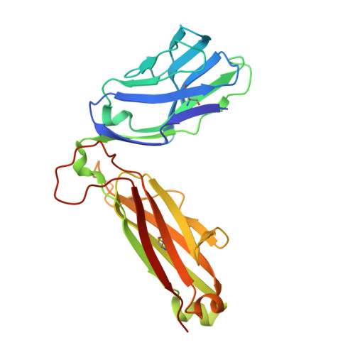
    5TEZ

  • PubMed Abstract: 

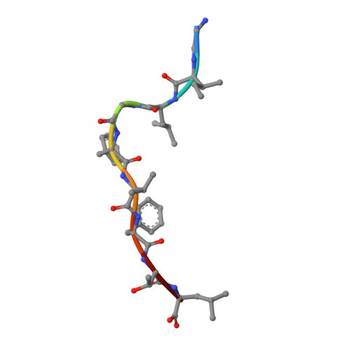
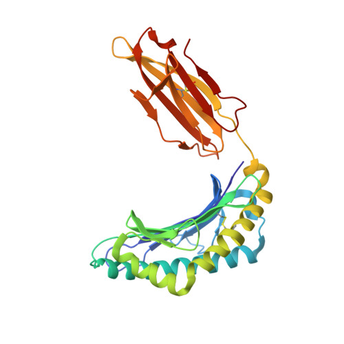
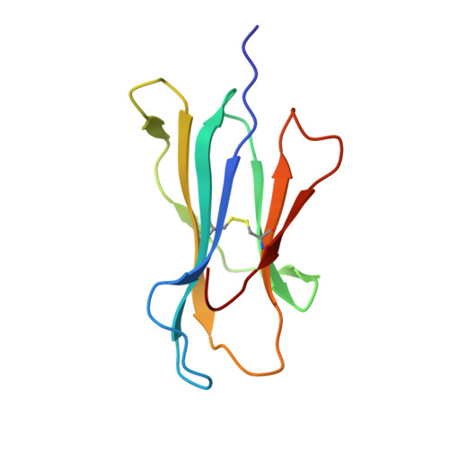
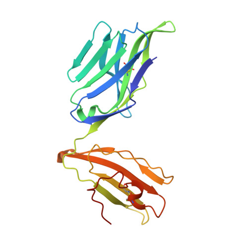
    Influenza A virus (IAV) causes an acute infection in humans that is normally eliminated by CD8 + cytotoxic T lymphocytes. Individuals expressing the MHC class I molecule HLA-A2 produce cytotoxic T lymphocytes bearing T-cell receptors (TCRs) that recognize the immunodominant IAV epitope GILGFVFTL (GIL). Most GIL-specific TCRs utilize α/β chain pairs encoded by the TRAV27/TRBV19 gene combination to recognize this relatively featureless peptide epitope (canonical TCRs). However, ∼40% of GIL-specific TCRs express a wide variety of other TRAV/TRBV combinations (non-canonical TCRs). To investigate the structural underpinnings of this remarkable diversity, we determined the crystal structure of a non-canonical GIL-specific TCR (F50) expressing the TRAV13-1/TRBV27 gene combination bound to GIL-HLA-A2 to 1.7 Å resolution. Comparison of the F50-GIL-HLA-A2 complex with the previously published complex formed by a canonical TCR (JM22) revealed that F50 and JM22 engage GIL-HLA-A2 in markedly different orientations. These orientations are distinguished by crossing angles of TCR to peptide-MHC of 29° for F50 versus 69° for JM22 and by a focus by F50 on the C terminus rather than the center of the MHC α1 helix for JM22. In addition, F50, unlike JM22, uses a tryptophan instead of an arginine to fill a critical notch between GIL and the HLA-A2 α2 helix. The F50-GIL-HLA-A2 complex shows that there are multiple structurally distinct solutions to recognizing an identical peptide-MHC ligand with sufficient affinity to elicit a broad anti-IAV response that protects against viral escape and T-cell clonal loss.


  • Organizational Affiliation
    • From the University of Maryland Institute for Bioscience and Biotechnology Research, W. M. Keck Laboratory for Structural Biology, Rockville, Maryland 20850.

Macromolecule Content 

  • Total Structure Weight: 95.47 kDa 
  • Atom Count: 7,466 
  • Modeled Residue Count: 826 
  • Deposited Residue Count: 835 
  • Unique protein chains: 5

Macromolecules

Find similar proteins by:  Sequence   |   3D Structure  
Entity ID: 1
MoleculeChains  Sequence LengthOrganismDetailsImage
GLY-ILE-LEU-GLY-PHE-VAL-PHE-THR-LEUA [auth C]9Influenza A virusMutation(s): 0 
UniProt
Find proteins for P03485 (Influenza A virus (strain A/Puerto Rico/8/1934 H1N1))
Explore P03485 
Go to UniProtKB:  P03485
Entity Groups
UniProt GroupP03485
Sequence Annotations
Expand
Reference Sequence
Find similar proteins by:|  3D Structure
Entity ID: 2
MoleculeChains  Sequence LengthOrganismDetailsImage
HLA class I histocompatibility antigen, A-2 alpha chainB [auth A]275Homo sapiensMutation(s): 0 
Gene Names: HLA-AHLAA
UniProt & NIH Common Fund Data Resources
Find proteins for P04439 (Homo sapiens)
Explore P04439 
Go to UniProtKB:  P04439
PHAROS:  P04439
GTEx:  ENSG00000206503 
Entity Groups
Sequence Clusters30% Identity50% Identity70% Identity90% Identity95% Identity100% Identity
UniProt GroupP04439
Sequence Annotations
Expand
Reference Sequence
Find similar proteins by:|  3D Structure
Entity ID: 3
MoleculeChains  Sequence LengthOrganismDetailsImage
Beta-2-microglobulinC [auth B]100Homo sapiensMutation(s): 0 
Gene Names: B2MCDABP0092HDCMA22P
UniProt & NIH Common Fund Data Resources
Find proteins for P61769 (Homo sapiens)
Explore P61769 
Go to UniProtKB:  P61769
PHAROS:  P61769
GTEx:  ENSG00000166710 
Entity Groups
Sequence Clusters30% Identity50% Identity70% Identity90% Identity95% Identity100% Identity
UniProt GroupP61769
Sequence Annotations
Expand
Reference Sequence
Find similar proteins by:|  3D Structure
Entity ID: 4
MoleculeChains  Sequence LengthOrganismDetailsImage
TCR F50 alpha chainD [auth I]208Homo sapiensMutation(s): 0 
Find similar proteins by:|  3D Structure
Entity ID: 5
MoleculeChains  Sequence LengthOrganismDetailsImage
TCR F50 beta chainE [auth J]243Homo sapiensMutation(s): 0 

Experimental Data & Validation

Experimental Data

  • Method: X-RAY DIFFRACTION
  • Resolution: 1.70 Å
  • R-Value Free:  0.224 (Depositor), 0.226 (DCC) 
  • R-Value Work:  0.190 (Depositor), 0.193 (DCC) 
  • R-Value Observed: 0.191 (Depositor) 
Space Group: P 1 21 1
Unit Cell:
Length ( Å )Angle ( ˚ )
a = 66.266α = 90
b = 71.053β = 96.22
c = 100.693γ = 90
Software Package:
Software NamePurpose
Aimlessdata scaling
PHENIXrefinement
PDB_EXTRACTdata extraction
HKL-2000data reduction
PHASERphasing

Structure Validation

View Full Validation Report


Entry History 

& Funding Information

Deposition Data


Funding OrganizationLocationGrant Number
National Institutes of Health/National Institute Of Allergy and Infectious Diseases (NIH/NIAID)United StatesAI036900

Revision History  (Full details and data files)

  • Version 1.0: 2017-09-27
    Type: Initial release
  • Version 1.1: 2017-10-04
    Changes: Database references
  • Version 1.2: 2017-11-22
    Changes: Database references
  • Version 1.3: 2019-12-11
    Changes: Author supporting evidence
  • Version 1.4: 2023-10-04
    Changes: Data collection, Database references, Refinement description
  • Version 1.5: 2024-11-13
    Changes: Structure summary