5TA0 | pdb_00005ta0

Crystal structure of BuGH86E322Q in complex with neoagarooctaose


Experimental Data Snapshot

  • Method: X-RAY DIFFRACTION
  • Resolution: 1.40 Å
  • R-Value Free: 
    0.162 (Depositor), 0.169 (DCC) 
  • R-Value Work: 
    0.142 (Depositor), 0.151 (DCC) 
  • R-Value Observed: 
    0.143 (Depositor) 

wwPDB Validation 3D Report Full Report

Validation slider image for 5TA0

This is version 2.1 of the entry. See complete history

Literature

Molecular basis of an agarose metabolic pathway acquired by a human intestinal symbiont.

Pluvinage, B.Grondin, J.M.Amundsen, C.Klassen, L.Moote, P.E.Xiao, Y.Thomas, D.Pudlo, N.A.Anele, A.Martens, E.C.Inglis, G.D.Uwiera, R.E.R.Boraston, A.B.Abbott, D.W.

(2018) Nat Commun 9: 1043-1043

  • DOI: https://doi.org/10.1038/s41467-018-03366-x
  • Primary Citation Related Structures: 
    5T98, 5T99, 5T9A, 5T9G, 5T9X, 5TA0, 5TA1, 5TA5, 5TA7, 5TA9

  • PubMed Abstract: 

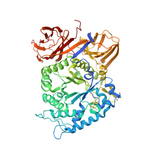
    In red algae, the most abundant principal cell wall polysaccharides are mixed galactan agars, of which agarose is a common component. While bioconversion of agarose is predominantly catalyzed by bacteria that live in the oceans, agarases have been discovered in microorganisms that inhabit diverse terrestrial ecosystems, including human intestines. Here we comprehensively define the structure-function relationship of the agarolytic pathway from the human intestinal bacterium Bacteroides uniformis (Bu) NP1. Using recombinant agarases from Bu NP1 to completely depolymerize agarose, we demonstrate that a non-agarolytic Bu strain can grow on GAL released from agarose. This relationship underscores that rare nutrient utilization by intestinal bacteria is facilitated by the acquisition of highly specific enzymes that unlock inaccessible carbohydrate resources contained within unusual polysaccharides. Intriguingly, the agarolytic pathway is differentially distributed throughout geographically distinct human microbiomes, reflecting a complex historical context for agarose consumption by human beings.


  • Organizational Affiliation
    • Department of Biochemistry and Microbiology, University of Victoria, PO Box 3055 STN CSC, Victoria, BC, V8W 3P6, Canada.

Macromolecule Content 

  • Total Structure Weight: 153.91 kDa 
  • Atom Count: 12,263 
  • Modeled Residue Count: 1,241 
  • Deposited Residue Count: 1,298 
  • Unique protein chains: 1

Macromolecules

Find similar proteins by:|  3D Structure
Entity ID: 1
MoleculeChains  Sequence LengthOrganismDetailsImage
Glycoside Hydrolase
A, B
649Bacteroides uniformisMutation(s): 0 
UniProt
Find proteins for A0A2D0TCD2 (Bacteroides uniformis)
Explore A0A2D0TCD2 
Go to UniProtKB:  A0A2D0TCD2
Entity Groups
Sequence Clusters30% Identity50% Identity70% Identity90% Identity95% Identity100% Identity
UniProt GroupA0A2D0TCD2
Sequence Annotations
Expand
Reference Sequence

Oligosaccharides

Help  
Entity ID: 2
MoleculeChains Length2D Diagram GlycosylationD Interactions
beta-D-galactopyranose-(1-4)-3,6-anhydro-alpha-L-galactopyranose-(1-3)-beta-D-galactopyranose-(1-4)-3,6-anhydro-alpha-L-galactopyranose-(1-3)-beta-D-galactopyranose
C
5N/A
Glycosylation Resources
GlyTouCan: G81067MN
GlyCosmos: G81067MN

Small Molecules

Ligands 4 Unique
IDChains Name / Formula / InChI Key2D Diagram3D Interactions
SO4

Query on SO4


Download:Ideal Coordinates CCD File
D [auth A]
E [auth A]
EA [auth B]
F [auth A]
FA [auth B]
D [auth A],
E [auth A],
EA [auth B],
F [auth A],
FA [auth B],
G [auth A],
GA [auth B],
HA [auth B]
SULFATE ION
O4 S
QAOWNCQODCNURD-UHFFFAOYSA-L
GOL

Query on GOL


Download:Ideal Coordinates CCD File
AA [auth A]
BA [auth A]
VA [auth B]
WA [auth B]
XA [auth B]
AA [auth A],
BA [auth A],
VA [auth B],
WA [auth B],
XA [auth B],
Y [auth A],
YA [auth B],
Z [auth A],
ZA [auth B]
GLYCEROL
C3 H8 O3
PEDCQBHIVMGVHV-UHFFFAOYSA-N
EDO

Query on EDO


Download:Ideal Coordinates CCD File
H [auth A]
I [auth A]
IA [auth B]
J [auth A]
JA [auth B]
H [auth A],
I [auth A],
IA [auth B],
J [auth A],
JA [auth B],
K [auth A],
KA [auth B],
L [auth A],
LA [auth B],
M [auth A],
MA [auth B],
N [auth A],
NA [auth B],
O [auth A],
OA [auth B],
P [auth A],
PA [auth B],
Q [auth A],
QA [auth B],
R [auth A],
RA [auth B],
S [auth A],
SA [auth B],
T [auth A],
TA [auth B],
U [auth A],
UA [auth B],
V [auth A],
W [auth A],
X [auth A]
1,2-ETHANEDIOL
C2 H6 O2
LYCAIKOWRPUZTN-UHFFFAOYSA-N
CA

Query on CA


Download:Ideal Coordinates CCD File
AB [auth B],
BB [auth B],
CA [auth A],
DA [auth A]
CALCIUM ION
Ca
BHPQYMZQTOCNFJ-UHFFFAOYSA-N

Experimental Data & Validation

Experimental Data

  • Method: X-RAY DIFFRACTION
  • Resolution: 1.40 Å
  • R-Value Free:  0.162 (Depositor), 0.169 (DCC) 
  • R-Value Work:  0.142 (Depositor), 0.151 (DCC) 
  • R-Value Observed: 0.143 (Depositor) 
Space Group: P 1
Unit Cell:
Length ( Å )Angle ( ˚ )
a = 60.719α = 85.87
b = 73.54β = 86.2
c = 83.19γ = 71.87
Software Package:
Software NamePurpose
MOSFLMdata reduction
SCALAdata scaling
PHASERphasing
REFMACrefinement
PDB_EXTRACTdata extraction

Structure Validation

View Full Validation Report


Entry History 

Deposition Data

Revision History  (Full details and data files)

  • Version 1.0: 2017-09-13
    Type: Initial release
  • Version 1.1: 2019-03-27
    Changes: Data collection, Database references
  • Version 2.0: 2020-07-29
    Type: Remediation
    Reason: Carbohydrate remediation
    Changes: Atomic model, Data collection, Derived calculations, Structure summary
  • Version 2.1: 2024-11-20
    Changes: Data collection, Database references, Derived calculations, Structure summary