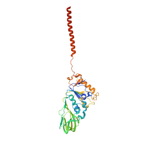
5T96

Crystal structure of the infectious salmon anemia virus (ISAV) HE viral receptor complex


Experimental Data Snapshot

  • Method: X-RAY DIFFRACTION
  • Resolution: 2.00 Å
  • R-Value Free: 0.183 
  • R-Value Work: 0.139 
  • R-Value Observed: 0.139 

Starting Model: experimental
View more details

wwPDB Validation   3D Report Full Report


Ligand Structure Quality Assessment 


This is version 2.2 of the entry. See complete history


Literature

Structure of the infectious salmon anemia virus receptor complex illustrates a unique binding strategy for attachment.

Cook, J.D.Sultana, A.Lee, J.E.

(2017) Proc Natl Acad Sci U S A 114: E2929-E2936

  • DOI: https://doi.org/10.1073/pnas.1617993114
  • Primary Citation of Related Structures:  
    5T96, 5T9Y

  • PubMed Abstract: 

    Orthomyxoviruses are an important family of RNA viruses, which include the various influenza viruses. Despite global efforts to eradicate orthomyxoviral pathogens, these infections remain pervasive. One such orthomyxovirus, infectious salmon anemia virus (ISAV), spreads easily throughout farmed and wild salmonids, constituting a significant economic burden. ISAV entry requires the interplay of the virion-attached hemagglutinin-esterase and fusion glycoproteins. Preventing infections will rely on improved understanding of ISAV entry. Here, we present the crystal structures of ISAV hemagglutinin-esterase unbound and complexed with receptor. Several distinctive features observed in ISAV HE are not seen in any other viral glycoprotein. The structures reveal a unique mode of receptor binding that is dependent on the oligomeric assembly of hemagglutinin-esterase. Importantly, ISAV hemagglutinin-esterase receptor engagement does not initiate conformational rearrangements, suggesting a distinct viral entry mechanism. This work improves our understanding of ISAV pathogenesis and expands our knowledge on the overall diversity of viral glycoprotein-mediated entry mechanisms. Finally, it provides an atomic-resolution model of the primary neutralizing antigen critical for vaccine development.


  • Organizational Affiliation

    Department of Laboratory Medicine and Pathobiology, Faculty of Medicine, University of Toronto, Toronto, ON M5S 1A8, Canada.


Macromolecules
Find similar proteins by:  (by identity cutoff)  |  3D Structure
Entity ID: 1
MoleculeChains Sequence LengthOrganismDetailsImage
HE protein
A, B, C, D, E
A, B, C, D, E, F
342Isavirus salarisMutation(s): 1 
Gene Names: HE
UniProt
Find proteins for Q9J0Y0 (Isavirus salaris)
Explore Q9J0Y0 
Go to UniProtKB:  Q9J0Y0
Entity Groups  
Sequence Clusters30% Identity50% Identity70% Identity90% Identity95% Identity100% Identity
UniProt GroupQ9J0Y0
Sequence Annotations
Expand
  • Reference Sequence
Small Molecules
Ligands 4 Unique
IDChains Name / Formula / InChI Key2D Diagram3D Interactions
79J
Query on 79J

Download Ideal Coordinates CCD File 
I [auth A]
IA [auth D]
P [auth B]
SA [auth E]
XA [auth F]
I [auth A],
IA [auth D],
P [auth B],
SA [auth E],
XA [auth F],
Z [auth C]
4-O-acetyl-5-acetamido-3,5-dideoxy-L-glycero-alpha-D-galacto-non-2-ulopyranosonic acid
C13 H21 N O10
LVBIMVQYUKOENY-DQBXORCPSA-N
GOL
Query on GOL

Download Ideal Coordinates CCD File 
AA [auth C]
J [auth A]
JA [auth D]
KA [auth D]
Q [auth B]
AA [auth C],
J [auth A],
JA [auth D],
KA [auth D],
Q [auth B],
R [auth B],
TA [auth E],
YA [auth F]
GLYCEROL
C3 H8 O3
PEDCQBHIVMGVHV-UHFFFAOYSA-N
FMT
Query on FMT

Download Ideal Coordinates CCD File 
AB [auth F]
BA [auth C]
BB [auth F]
CA [auth C]
DA [auth C]
AB [auth F],
BA [auth C],
BB [auth F],
CA [auth C],
DA [auth C],
EA [auth C],
FA [auth C],
K [auth A],
L [auth A],
LA [auth D],
M [auth A],
MA [auth D],
N [auth A],
NA [auth D],
OA [auth D],
PA [auth D],
QA [auth D],
RA [auth D],
S [auth B],
T [auth B],
U [auth B],
UA [auth E],
V [auth B],
VA [auth E],
W [auth B],
X [auth B],
Y [auth C],
ZA [auth F]
FORMIC ACID
C H2 O2
BDAGIHXWWSANSR-UHFFFAOYSA-N
MG
Query on MG

Download Ideal Coordinates CCD File 
G [auth A]
GA [auth D]
H [auth A]
HA [auth D]
O [auth B]
G [auth A],
GA [auth D],
H [auth A],
HA [auth D],
O [auth B],
WA [auth F]
MAGNESIUM ION
Mg
JLVVSXFLKOJNIY-UHFFFAOYSA-N
Experimental Data & Validation

Experimental Data

  • Method: X-RAY DIFFRACTION
  • Resolution: 2.00 Å
  • R-Value Free: 0.183 
  • R-Value Work: 0.139 
  • R-Value Observed: 0.139 
  • Space Group: P 1
Unit Cell:
Length ( Å )Angle ( ˚ )
a = 82.48α = 97.14
b = 87.568β = 113.5
c = 95.534γ = 114.68
Software Package:
Software NamePurpose
PHENIXrefinement
Aimlessdata scaling
XDSdata reduction
PDB_EXTRACTdata extraction
Cootmodel building
PHASERphasing

Structure Validation

View Full Validation Report


Ligand Structure Quality Assessment 


Entry History 

Deposition Data

Revision History  (Full details and data files)

  • Version 1.0: 2017-03-22
    Type: Initial release
  • Version 1.1: 2017-04-05
    Changes: Database references
  • Version 1.2: 2017-04-19
    Changes: Database references
  • Version 2.0: 2020-07-29
    Type: Remediation
    Reason: Carbohydrate remediation
    Changes: Atomic model, Data collection, Derived calculations, Structure summary
  • Version 2.1: 2023-10-04
    Changes: Data collection, Database references, Refinement description, Structure summary
  • Version 2.2: 2024-11-13
    Changes: Structure summary