5T8O | pdb_00005t8o

Crystal structure of murine NF-kappaB inducing kinase (NIK) bound to Imidazobenzoxepin Compound 3


Experimental Data Snapshot

  • Method: X-RAY DIFFRACTION
  • Resolution: 2.41 Å
  • R-Value Free: 
    0.256 (Depositor), 0.258 (DCC) 
  • R-Value Work: 
    0.201 (Depositor), 0.204 (DCC) 
  • R-Value Observed: 
    0.207 (Depositor) 

wwPDB Validation 3D Report Full Report

Validation slider image for 5T8O

Ligand Structure Quality Assessment 


This is version 1.3 of the entry. See complete history

Literature

Structure-Based Design of Tricyclic NF-kappa B Inducing Kinase (NIK) Inhibitors That Have High Selectivity over Phosphoinositide-3-kinase (PI3K).

Castanedo, G.M.Blaquiere, N.Beresini, M.Bravo, B.Brightbill, H.Chen, J.Cui, H.F.Eigenbrot, C.Everett, C.Feng, J.Godemann, R.Gogol, E.Hymowitz, S.Johnson, A.Kayagaki, N.Kohli, P.B.Knuppel, K.Kraemer, J.Kruger, S.Loke, P.McEwan, P.Montalbetti, C.Roberts, D.A.Smith, M.Steinbacher, S.Sujatha-Bhaskar, S.Takahashi, R.Wang, X.Wu, L.C.Zhang, Y.Staben, S.T.

(2017) J Med Chem 60: 627-640

  • DOI: https://doi.org/10.1021/acs.jmedchem.6b01363
  • Primary Citation Related Structures: 
    5T8F, 5T8O, 5T8P, 5T8Q

  • PubMed Abstract: 

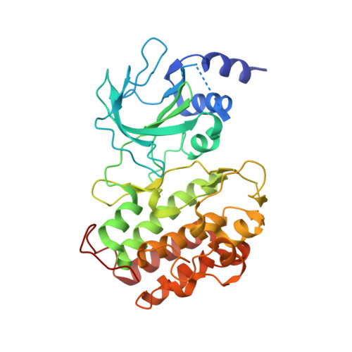
    We report here structure-guided optimization of a novel series of NF-κB inducing kinase (NIK) inhibitors. Starting from a modestly potent, low molecular weight lead, activity was improved by designing a type 11/2 binding mode that accessed a back pocket past the methionine-471 gatekeeper. Divergent binding modes in NIK and PI3K were exploited to dampen PI3K inhibition while maintaining NIK inhibition within these series. Potent compounds were discovered that selectively inhibit the nuclear translocation of NF-κB2 (p52/REL-B) but not canonical NF-κB1 (REL-A/p50).


  • Organizational Affiliation
    • Genentech, Inc. 1 DNA Way, South San Francisco, California 94080, United States.

Macromolecule Content 

  • Total Structure Weight: 78.38 kDa 
  • Atom Count: 5,462 
  • Modeled Residue Count: 652 
  • Deposited Residue Count: 698 
  • Unique protein chains: 1

Macromolecules

Find similar proteins by:|  3D Structure
Entity ID: 1
MoleculeChains  Sequence LengthOrganismDetailsImage
Mitogen-activated protein kinase kinase kinase 14
A, B
349Mus musculusMutation(s): 0 
Gene Names: Map3k14Nik
EC: 2.7.11.25
UniProt
Find proteins for Q9WUL6 (Mus musculus)
Explore Q9WUL6 
Go to UniProtKB:  Q9WUL6
Entity Groups
Sequence Clusters30% Identity50% Identity70% Identity90% Identity95% Identity100% Identity
UniProt GroupQ9WUL6
Sequence Annotations
Expand
Reference Sequence

Small Molecules

Ligands 2 Unique
IDChains Name / Formula / InChI Key2D Diagram3D Interactions
76Z

Query on 76Z


Download:Ideal Coordinates CCD File
G [auth A],
K [auth B]
10-(3-methyl-3-oxidanyl-but-1-ynyl)-5,6-dihydroimidazo[1,2-d][1,4]benzoxazepine-2-carboxamide
C17 H17 N3 O3
CMHKURIYUXPNNR-UHFFFAOYSA-N
SO4

Query on SO4


Download:Ideal Coordinates CCD File
C [auth A]
D [auth A]
E [auth A]
F [auth A]
H [auth B]
C [auth A],
D [auth A],
E [auth A],
F [auth A],
H [auth B],
I [auth B],
J [auth B]
SULFATE ION
O4 S
QAOWNCQODCNURD-UHFFFAOYSA-L

Experimental Data & Validation

Experimental Data

  • Method: X-RAY DIFFRACTION
  • Resolution: 2.41 Å
  • R-Value Free:  0.256 (Depositor), 0.258 (DCC) 
  • R-Value Work:  0.201 (Depositor), 0.204 (DCC) 
  • R-Value Observed: 0.207 (Depositor) 
Space Group: P 43
Unit Cell:
Length ( Å )Angle ( ˚ )
a = 143.95α = 90
b = 143.95β = 90
c = 45.47γ = 90
Software Package:
Software NamePurpose
REFMACrefinement
MOSFLMdata reduction
SCALAdata scaling
PHASERphasing

Structure Validation

View Full Validation Report


Ligand Structure Quality Assessment 


Entry History 

Deposition Data

Revision History  (Full details and data files)

  • Version 1.0: 2017-01-11
    Type: Initial release
  • Version 1.1: 2017-01-25
    Changes: Database references
  • Version 1.2: 2017-02-08
    Changes: Database references
  • Version 1.3: 2024-05-08
    Changes: Data collection, Database references