5T76 | pdb_00005t76

A fragment of a human tRNA synthetase


Experimental Data Snapshot

  • Method: X-RAY DIFFRACTION
  • Resolution: 2.00 Å
  • R-Value Free: 
    0.258 (Depositor), 0.255 (DCC) 
  • R-Value Work: 
    0.215 (Depositor), 0.216 (DCC) 

wwPDB Validation 3D Report Full Report

Validation slider image for 5T76

This is version 1.2 of the entry. See complete history

Literature

Two crystal structures reveal design for repurposing the C-Ala domain of human AlaRS.

Sun, L.Song, Y.Blocquel, D.Yang, X.L.Schimmel, P.

(2016) Proc Natl Acad Sci U S A 113: 14300-14305

  • DOI: https://doi.org/10.1073/pnas.1617316113
  • Primary Citation Related Structures: 
    5T5S, 5T76

  • PubMed Abstract: 

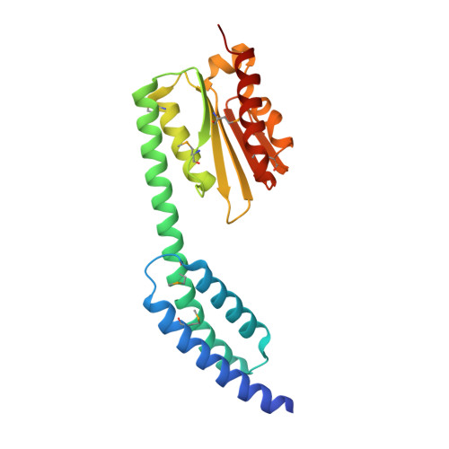
    The 20 aminoacyl tRNA synthetases (aaRSs) couple each amino acid to their cognate tRNAs. During evolution, 19 aaRSs expanded by acquiring novel noncatalytic appended domains, which are absent from bacteria and many lower eukaryotes but confer extracellular and nuclear functions in higher organisms. AlaRS is the single exception, with an appended C-terminal domain (C-Ala) that is conserved from prokaryotes to humans but with a wide sequence divergence. In human cells, C-Ala is also a splice variant of AlaRS. Crystal structures of two forms of human C-Ala, and small-angle X-ray scattering of AlaRS, showed that the large sequence divergence of human C-Ala reshaped C-Ala in a way that changed the global architecture of AlaRS. This reshaping removes the role of C-Ala in prokaryotes for docking tRNA and instead repurposes it to form a dimer interface presenting a DNA-binding groove. This groove cannot form with the bacterial ortholog. Direct DNA binding by human C-Ala, but not by bacterial C-Ala, was demonstrated. Thus, instead of acquiring a novel appended domain like other human aaRSs, which engendered novel functions, a new AlaRS architecture was created by diversifying a preexisting appended domain.


  • Organizational Affiliation
    • The Scripps Laboratories for tRNA Synthetase Research, The Scripps Research Institute, La Jolla, CA 92037.

Macromolecule Content 

  • Total Structure Weight: 23.75 kDa 
  • Atom Count: 1,740 
  • Modeled Residue Count: 208 
  • Deposited Residue Count: 215 
  • Unique protein chains: 1

Macromolecules

Find similar proteins by:|  3D Structure
Entity ID: 1
MoleculeChains  Sequence LengthOrganismDetailsImage
Alanine--tRNA ligase, cytoplasmic215Homo sapiensMutation(s): 0 
Gene Names: AARS
EC: 6.1.1.7 (PDB Primary Data), 6 (UniProt)
UniProt & NIH Common Fund Data Resources
Find proteins for P49588 (Homo sapiens)
Explore P49588 
Go to UniProtKB:  P49588
PHAROS:  P49588
GTEx:  ENSG00000090861 
Entity Groups
Sequence Clusters30% Identity50% Identity70% Identity90% Identity95% Identity100% Identity
UniProt GroupP49588
Sequence Annotations
Expand
Reference Sequence

Small Molecules

Modified Residues  1 Unique
IDChains TypeFormula2D DiagramParent
MSE
Query on MSE
A
L-PEPTIDE LINKINGC5 H11 N O2 SeMET

Experimental Data & Validation

Experimental Data

  • Method: X-RAY DIFFRACTION
  • Resolution: 2.00 Å
  • R-Value Free:  0.258 (Depositor), 0.255 (DCC) 
  • R-Value Work:  0.215 (Depositor), 0.216 (DCC) 
Space Group: P 1 21 1
Unit Cell:
Length ( Å )Angle ( ˚ )
a = 41.645α = 90
b = 38.449β = 90.5
c = 62.471γ = 90
Software Package:
Software NamePurpose
REFMACrefinement
HKL-2000data scaling
PHENIXphasing
HKL-2000data reduction

Structure Validation

View Full Validation Report


Entry History 

Deposition Data

Revision History  (Full details and data files)

  • Version 1.0: 2016-11-30
    Type: Initial release
  • Version 1.1: 2017-01-04
    Changes: Database references
  • Version 1.2: 2024-11-13
    Changes: Data collection, Database references, Structure summary