5T6S | pdb_00005t6s

Crystal structure of the A/Shanghai/2/2013 (H7N9) influenza virus hemagglutinin in complex with the antiviral drug arbidol


Experimental Data Snapshot

  • Method: X-RAY DIFFRACTION
  • Resolution: 2.36 Å
  • R-Value Free: 
    0.250 (Depositor), 0.251 (DCC) 
  • R-Value Work: 
    0.204 (Depositor), 0.207 (DCC) 
  • R-Value Observed: 
    0.206 (Depositor) 

Starting Model: experimental
View more details

wwPDB Validation 3D Report Full Report

Validation slider image for 5T6S

Ligand Structure Quality Assessment 


This is version 2.2 of the entry. See complete history

Literature

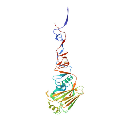
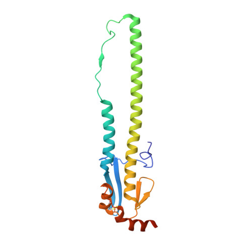
Structural basis of influenza virus fusion inhibition by the antiviral drug Arbidol.

Kadam, R.U.Wilson, I.A.

(2017) Proc Natl Acad Sci U S A 114: 206-214

  • DOI: https://doi.org/10.1073/pnas.1617020114
  • Primary Citation Related Structures: 
    5T6N, 5T6S

  • PubMed Abstract: 

    The broad-spectrum antiviral drug Arbidol shows efficacy against influenza viruses by targeting the hemagglutinin (HA) fusion machinery. However, the structural basis of the mechanism underlying fusion inhibition by Arbidol has remained obscure, thereby hindering its further development as a specific and optimized influenza therapeutic. We determined crystal structures of Arbidol in complex with influenza virus HA from pandemic 1968 H3N2 and recent 2013 H7N9 viruses. Arbidol binds in a hydrophobic cavity in the HA trimer stem at the interface between two protomers. This cavity is distal to the conserved epitope targeted by broadly neutralizing stem antibodies and is ∼16 Å from the fusion peptide. Arbidol primarily makes hydrophobic interactions with the binding site but also induces some conformational rearrangements to form a network of inter- and intraprotomer salt bridges. By functioning as molecular glue, Arbidol stabilizes the prefusion conformation of HA that inhibits the large conformational rearrangements associated with membrane fusion in the low pH of the endosome. This unique binding mode compared with the small-molecule inhibitors of other class I fusion proteins enhances our understanding of how small molecules can function as fusion inhibitors and guides the development of broad-spectrum therapeutics against influenza virus.


  • Organizational Affiliation
    • Department of Integrative Structural and Computational Biology, The Scripps Research Institute, La Jolla, CA 92037.

Macromolecule Content 

  • Total Structure Weight: 346.44 kDa 
  • Atom Count: 24,445 
  • Modeled Residue Count: 2,922 
  • Deposited Residue Count: 3,024 
  • Unique protein chains: 2

Macromolecules

Find similar proteins by:|  3D Structure
Entity ID: 1
MoleculeChains  Sequence LengthOrganismDetailsImage
Hemagglutinin HA1
A, C, E, G, I
A, C, E, G, I, K
321Influenza A virus (A/Shanghai/02/2013(H7N9))Mutation(s): 0 
Gene Names: HA
UniProt
Find proteins for R4NN21 (Influenza A virus)
Explore R4NN21 
Go to UniProtKB:  R4NN21
Entity Groups
Sequence Clusters30% Identity50% Identity70% Identity90% Identity95% Identity100% Identity
UniProt GroupR4NN21
Glycosylation
Glycosylation Sites: 2
Sequence Annotations
Expand
Reference Sequence
Find similar proteins by:|  3D Structure
Entity ID: 2
MoleculeChains  Sequence LengthOrganismDetailsImage
Hemagglutinin HA2
B, D, F, H, J
B, D, F, H, J, L
183Influenza A virus (A/Shanghai/02/2013(H7N9))Mutation(s): 0 
Gene Names: HA
UniProt
Find proteins for R4NN21 (Influenza A virus)
Explore R4NN21 
Go to UniProtKB:  R4NN21
Entity Groups
Sequence Clusters30% Identity50% Identity70% Identity90% Identity95% Identity100% Identity
UniProt GroupR4NN21
Glycosylation
Glycosylation Sites: 2
Sequence Annotations
Expand
Reference Sequence

Oligosaccharides

Help  
Entity ID: 3
MoleculeChains Length2D Diagram GlycosylationD Interactions
beta-D-mannopyranose-(1-4)-2-acetamido-2-deoxy-beta-D-glucopyranose-(1-4)-2-acetamido-2-deoxy-beta-D-glucopyranose
M, N, Q, R, S
3N-Glycosylation
Glycosylation Resources
GlyTouCan: G15407YE
GlyCosmos: G15407YE
GlyGen: G15407YE
Entity ID: 4
MoleculeChains Length2D Diagram GlycosylationD Interactions
2-acetamido-2-deoxy-beta-D-glucopyranose-(1-4)-2-acetamido-2-deoxy-beta-D-glucopyranose
O, P
2N-Glycosylation
Glycosylation Resources
GlyTouCan: G42666HT
GlyCosmos: G42666HT
GlyGen: G42666HT

Small Molecules

Ligands 3 Unique
IDChains Name / Formula / InChI Key2D Diagram3D Interactions
75U

Query on 75U


Download:Ideal Coordinates CCD File
DA [auth F]
FA [auth H]
IA [auth J]
NA [auth L]
V [auth B]
DA [auth F],
FA [auth H],
IA [auth J],
NA [auth L],
V [auth B],
W [auth B]
ethyl 6-bromo-4-[(dimethylamino)methyl]-5-hydroxy-1-methyl-2-[(phenylsulfanyl)methyl]-1H-indole-3-carboxylate
C22 H25 Br N2 O3 S
KCFYEAOKVJSACF-UHFFFAOYSA-N
NAG

Query on NAG


Download:Ideal Coordinates CCD File
AA [auth D]
BA [auth D]
CA [auth E]
EA [auth F]
GA [auth H]
AA [auth D],
BA [auth D],
CA [auth E],
EA [auth F],
GA [auth H],
HA [auth I],
JA [auth J],
KA [auth J],
MA [auth K],
OA [auth L],
T [auth A],
U [auth A],
X [auth B],
Y [auth B],
Z [auth C]
2-acetamido-2-deoxy-beta-D-glucopyranose
C8 H15 N O6
OVRNDRQMDRJTHS-FMDGEEDCSA-N
NA

Query on NA


Download:Ideal Coordinates CCD File
LA [auth J]SODIUM ION
Na
FKNQFGJONOIPTF-UHFFFAOYSA-N

Experimental Data & Validation

Experimental Data

  • Method: X-RAY DIFFRACTION
  • Resolution: 2.36 Å
  • R-Value Free:  0.250 (Depositor), 0.251 (DCC) 
  • R-Value Work:  0.204 (Depositor), 0.207 (DCC) 
  • R-Value Observed: 0.206 (Depositor) 
Space Group: P 1 21 1
Unit Cell:
Length ( Å )Angle ( ˚ )
a = 67.419α = 90
b = 231.49β = 96.88
c = 127.85γ = 90
Software Package:
Software NamePurpose
PHENIXrefinement
Cootmodel building
HKL-2000data scaling
PHASERphasing
HKL-2000data reduction

Structure Validation

View Full Validation Report


Ligand Structure Quality Assessment 


Entry History 

Deposition Data

Revision History  (Full details and data files)

  • Version 1.0: 2017-01-04
    Type: Initial release
  • Version 1.1: 2017-01-25
    Changes: Database references
  • Version 2.0: 2020-07-29
    Type: Remediation
    Reason: Carbohydrate remediation
    Changes: Advisory, Atomic model, Data collection, Derived calculations, Structure summary
  • Version 2.1: 2023-10-04
    Changes: Data collection, Database references, Refinement description, Structure summary
  • Version 2.2: 2024-10-23
    Changes: Structure summary