


Crystal structure of Short-chain dehydrogenase/reductase SDR:Glucose/ribitol dehydrogenase from Brucella melitensis
Abendroth, J., Phan, J.N., Mayclin, S.J., Lorimer, D.D., Edwards, T.E.To be published.
Experimental Data Snapshot
Starting Model: experimental
View more details
Entity ID: 1 | |||||
|---|---|---|---|---|---|
| Molecule | Chains | Sequence Length | Organism | Details | Image |
| Short-chain dehydrogenase/reductase SDR:Glucose/ribitol dehydrogenase | 247 | Brucella abortus 2308 | Mutation(s): 0 Gene Names: BAB2_0029 EC: 1.1.1.100 | 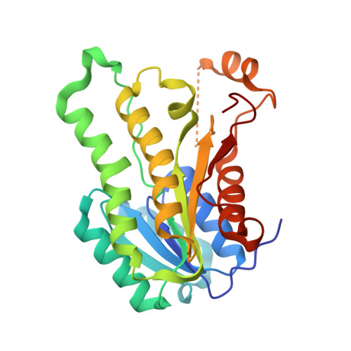 | |
UniProt | |||||
Entity Groups | |||||
| Sequence Clusters | 30% Identity50% Identity70% Identity90% Identity95% Identity100% Identity | ||||
| UniProt Group | Q2YL80 | ||||
Sequence AnnotationsExpand | |||||
Reference Sequence | |||||
| Ligands 3 Unique | |||||
|---|---|---|---|---|---|
| ID | Chains | Name / Formula / InChI Key | 2D Diagram | 3D Interactions | |
| NAD Download:Ideal Coordinates CCD File | E [auth A], G [auth B], J [auth C], L [auth D] | NICOTINAMIDE-ADENINE-DINUCLEOTIDE C21 H27 N7 O14 P2 BAWFJGJZGIEFAR-NNYOXOHSSA-N |  | ||
| MPD Download:Ideal Coordinates CCD File | H [auth B], I [auth C] | (4S)-2-METHYL-2,4-PENTANEDIOL C6 H14 O2 SVTBMSDMJJWYQN-YFKPBYRVSA-N |  | ||
| SO4 Download:Ideal Coordinates CCD File | F [auth B], K [auth C] | SULFATE ION O4 S QAOWNCQODCNURD-UHFFFAOYSA-L |  | ||
| Length ( Å ) | Angle ( ˚ ) |
|---|---|
| a = 64.85 | α = 90 |
| b = 108.15 | β = 103.12 |
| c = 65.66 | γ = 90 |
| Software Name | Purpose |
|---|---|
| XSCALE | data scaling |
| MOLREP | phasing |
| PHENIX | refinement |
| PDB_EXTRACT | data extraction |
| XDS | data scaling |
| ARP | model building |
| Coot | model building |
| XDS | data reduction |


