5T49 | pdb_00005t49

Crystal structure of SeMet derivative BhGH81


Experimental Data Snapshot

  • Method: X-RAY DIFFRACTION
  • Resolution: 2.50 Å
  • R-Value Free: 
    0.189 (Depositor), 0.189 (DCC) 
  • R-Value Work: 
    0.142 (Depositor), 0.142 (DCC) 
  • R-Value Observed: 
    0.144 (Depositor) 

wwPDB Validation 3D Report Full Report

Validation slider image for 5T49

This is version 1.3 of the entry. See complete history

Literature

Structural Analysis of a Family 81 Glycoside Hydrolase Implicates Its Recognition of beta-1,3-Glucan Quaternary Structure.

Pluvinage, B.Fillo, A.Massel, P.Boraston, A.B.

(2017) Structure 25: 1348-1359.e3

  • DOI: https://doi.org/10.1016/j.str.2017.06.019
  • Primary Citation Related Structures: 
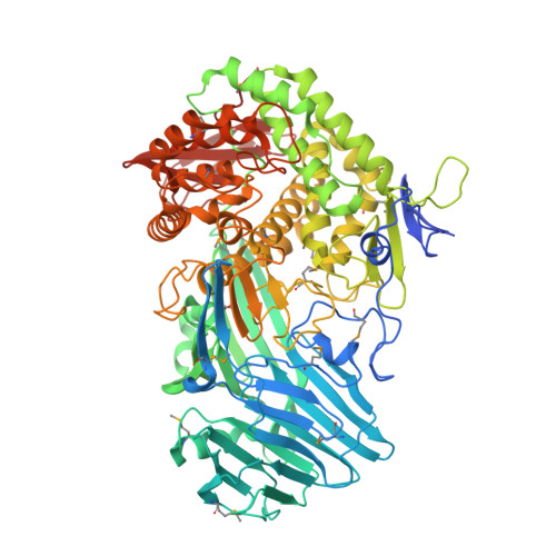
    5T49, 5T4A, 5T4C, 5T4G

  • PubMed Abstract: 

    Family 81 glycoside hydrolases (GHs), which are known to cleave β-1,3-glucans, are found in archaea, bacteria, eukaryotes, and viruses. Here we examine the structural and functional features of the GH81 catalytic module, BhGH81, from the Bacillus halodurans protein BH0236 to probe the molecular basis of β-1,3-glucan recognition and cleavage. BhGH81 displayed activity on laminarin, curdlan, and pachyman, but not scleroglucan; the enzyme also cleaved β-1,3-glucooligosaccharides as small as β-1,3-glucotriose. The crystal structures of BhGH81 in complex with various β-1,3-glucooligosaccharides revealed distorted sugars in the -1 catalytic subsite and an arrangement consistent with an inverting catalytic mechanism having a proposed conformational itinerary of 2 S 0 2,5 B 5 S 1 . Notably, the architecture of the catalytic site, location of an adjacent ancillary β-1,3-glucan binding site, and the surface properties of the enzyme indicate the likely ability to recognize the double and/or triple-helical quaternary structures adopted by β-1,3-glucans.


  • Organizational Affiliation
    • Department of Biochemistry and Microbiology, University of Victoria, Victoria, BC V8W 3P6, Canada.

Macromolecule Content 

  • Total Structure Weight: 90.86 kDa 
  • Atom Count: 6,465 
  • Modeled Residue Count: 752 
  • Deposited Residue Count: 785 
  • Unique protein chains: 1

Macromolecules

Find similar proteins by:|  3D Structure
Entity ID: 1
MoleculeChains  Sequence LengthOrganismDetailsImage
BH0236 protein785Halalkalibacterium halodurans C-125Mutation(s): 0 
Gene Names: BH0236
EC: 3.2.1.39
UniProt
Find proteins for Q9KG76 (Halalkalibacterium halodurans (strain ATCC BAA-125 / DSM 18197 / FERM 7344 / JCM 9153 / C-125))
Explore Q9KG76 
Go to UniProtKB:  Q9KG76
Entity Groups
Sequence Clusters30% Identity50% Identity70% Identity90% Identity95% Identity100% Identity
UniProt GroupQ9KG76
Sequence Annotations
Expand
Reference Sequence

Small Molecules

Ligands 2 Unique
IDChains Name / Formula / InChI Key2D Diagram3D Interactions
PO4

Query on PO4


Download:Ideal Coordinates CCD File
K [auth A]
L [auth A]
M [auth A]
N [auth A]
O [auth A]
K [auth A],
L [auth A],
M [auth A],
N [auth A],
O [auth A],
P [auth A],
Q [auth A],
R [auth A],
S [auth A]
PHOSPHATE ION
O4 P
NBIIXXVUZAFLBC-UHFFFAOYSA-K
EDO

Query on EDO


Download:Ideal Coordinates CCD File
B [auth A]
C [auth A]
D [auth A]
E [auth A]
F [auth A]
B [auth A],
C [auth A],
D [auth A],
E [auth A],
F [auth A],
G [auth A],
H [auth A],
I [auth A],
J [auth A]
1,2-ETHANEDIOL
C2 H6 O2
LYCAIKOWRPUZTN-UHFFFAOYSA-N
Modified Residues  1 Unique
IDChains TypeFormula2D DiagramParent
MSE
Query on MSE
A
L-PEPTIDE LINKINGC5 H11 N O2 SeMET

Experimental Data & Validation

Experimental Data

  • Method: X-RAY DIFFRACTION
  • Resolution: 2.50 Å
  • R-Value Free:  0.189 (Depositor), 0.189 (DCC) 
  • R-Value Work:  0.142 (Depositor), 0.142 (DCC) 
  • R-Value Observed: 0.144 (Depositor) 
Space Group: P 21 21 21
Unit Cell:
Length ( Å )Angle ( ˚ )
a = 65.54α = 90
b = 94.29β = 90
c = 159.17γ = 90
Software Package:
Software NamePurpose
MOSFLMdata reduction
SCALAdata scaling
PHASERphasing
SHARPphasing
REFMACrefinement
PDB_EXTRACTdata extraction

Structure Validation

View Full Validation Report


Entry History 

& Funding Information

Deposition Data


Funding OrganizationLocationGrant Number
Natural Sciences and Engineering Research Council (NSERC, Canada)Canada--

Revision History  (Full details and data files)

  • Version 1.0: 2017-06-28
    Type: Initial release
  • Version 1.1: 2019-03-27
    Changes: Author supporting evidence, Data collection, Database references
  • Version 1.2: 2020-01-08
    Changes: Author supporting evidence
  • Version 1.3: 2024-11-13
    Changes: Data collection, Database references, Structure summary