5T3F | pdb_00005t3f

hen egg-white lysozyme soaked with selenourea for 10 min

  • Classification: HYDROLASE
  • Organism(s): Gallus gallus
  • Mutation(s): No 

  • Deposited: 2016-08-25 Released: 2016-11-30 
  • Deposition Author(s): Luo, Z., Dauter, Z.
  • Funding Organization(s): National Institutes of Health/National Cancer Institute (NIH/NCI)

Experimental Data Snapshot

  • Method: X-RAY DIFFRACTION
  • Resolution: 1.45 Å
  • R-Value Free: 
    0.184 (Depositor), 0.186 (DCC) 
  • R-Value Work: 
    0.134 (Depositor), 0.137 (DCC) 
  • R-Value Observed: 
    0.136 (Depositor) 

wwPDB Validation 3D Report Full Report

Validation slider image for 5T3F

This is version 1.3 of the entry. See complete history

Literature

Selenourea: a convenient phasing vehicle for macromolecular X-ray crystal structures.

Luo, Z.

(2016) Sci Rep 6: 37123-37123

  • DOI: https://doi.org/10.1038/srep37123
  • Primary Citation Related Structures: 
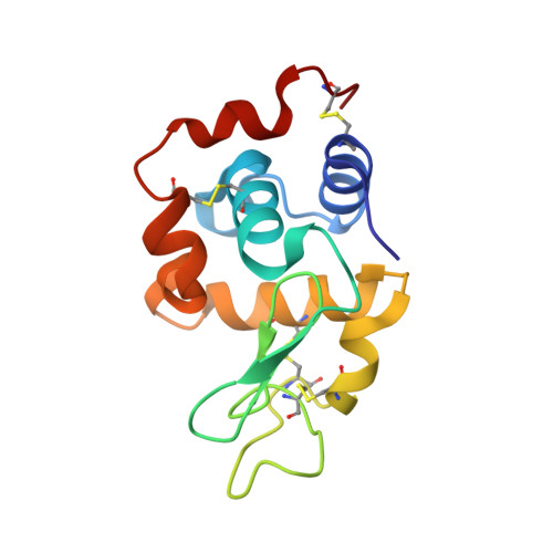
    5T3F, 5T3G, 5T3H, 5T3I, 5T3J, 5T3L

  • PubMed Abstract: 

    Majority of novel X-ray crystal structures of proteins are currently solved using the anomalous diffraction signal provided by selenium after incorporation of selenomethionine instead of natural methionine by genetic engineering methods. However, selenium can be inserted into protein crystals in the form of selenourea (SeC(NH 2 ) 2 ), by adding the crystalline powder of selenourea into mother liquor or cryo-solution with native crystals, in analogy to the classic procedure of heavy-atom derivatization. Selenourea is able to bind to reactive groups at the surface of macromolecules primarily through hydrogen bonds, where the selenium atom may serve as acceptor and amide groups as donors. Selenourea has different chemical properties than heavy-atom reagents and halide ions and provides a convenient way of phasing crystal structures of macromolecules.


  • Organizational Affiliation
    • Synchrotron Radiation Research Section, National Cancer Institute, Argonne National Laboratory, Argonne, 60439, USA.

Macromolecule Content 

  • Total Structure Weight: 15.44 kDa 
  • Atom Count: 1,162 
  • Modeled Residue Count: 129 
  • Deposited Residue Count: 129 
  • Unique protein chains: 1

Macromolecules

Find similar proteins by:|  3D Structure
Entity ID: 1
MoleculeChains  Sequence LengthOrganismDetailsImage
Lysozyme C129Gallus gallusMutation(s): 0 
EC: 3.2.1.17
UniProt
Find proteins for P00698 (Gallus gallus)
Explore P00698 
Go to UniProtKB:  P00698
Entity Groups
Sequence Clusters30% Identity50% Identity70% Identity90% Identity95% Identity100% Identity
UniProt GroupP00698
Sequence Annotations
Expand
Reference Sequence

Small Molecules

Ligands 1 Unique
IDChains Name / Formula / InChI Key2D Diagram3D Interactions
SEY

Query on SEY


Download:Ideal Coordinates CCD File
B [auth A]
C [auth A]
D [auth A]
E [auth A]
F [auth A]
B [auth A],
C [auth A],
D [auth A],
E [auth A],
F [auth A],
G [auth A],
H [auth A],
I [auth A],
J [auth A]
selenourea
C H4 N2 Se
IYKVLICPFCEZOF-UHFFFAOYSA-N

Experimental Data & Validation

Experimental Data

  • Method: X-RAY DIFFRACTION
  • Resolution: 1.45 Å
  • R-Value Free:  0.184 (Depositor), 0.186 (DCC) 
  • R-Value Work:  0.134 (Depositor), 0.137 (DCC) 
  • R-Value Observed: 0.136 (Depositor) 
Space Group: P 43 21 2
Unit Cell:
Length ( Å )Angle ( ˚ )
a = 77.37α = 90
b = 77.37β = 90
c = 37.306γ = 90
Software Package:
Software NamePurpose
REFMACrefinement
HKL-3000data reduction
HKL-3000data scaling
SHELXDphasing

Structure Validation

View Full Validation Report


Entry History 

& Funding Information

Deposition Data


Funding OrganizationLocationGrant Number
National Institutes of Health/National Cancer Institute (NIH/NCI)United States--

Revision History  (Full details and data files)

  • Version 1.0: 2016-11-30
    Type: Initial release
  • Version 1.1: 2017-09-27
    Changes: Author supporting evidence
  • Version 1.2: 2019-12-04
    Changes: Author supporting evidence, Derived calculations
  • Version 1.3: 2024-11-13
    Changes: Data collection, Database references, Structure summary