5T1I | pdb_00005t1i

CBX3 chromo shadow domain in complex with histone H3 peptide


Experimental Data Snapshot

  • Method: X-RAY DIFFRACTION
  • Resolution: 1.60 Å
  • R-Value Free: 
    0.239 (Depositor), 0.247 (DCC) 
  • R-Value Work: 
    0.194 (Depositor), 0.206 (DCC) 
  • R-Value Observed: 
    0.197 (Depositor) 

Starting Model: experimental
View more details

wwPDB Validation 3D Report Full Report

Validation slider image for 5T1I

This is version 1.5 of the entry. See complete history

Literature

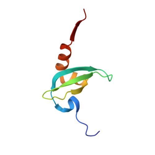
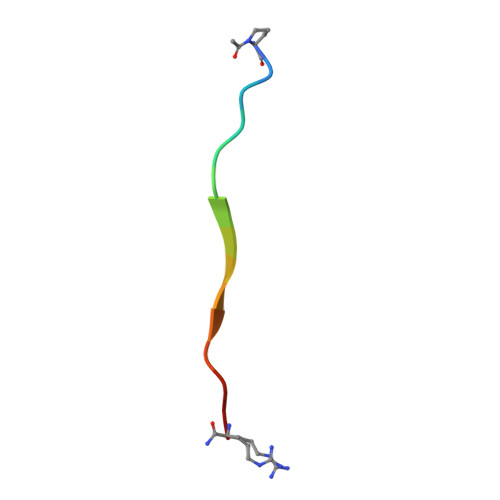
Peptide recognition by heterochromatin protein 1 (HP1) chromoshadow domains revisited: Plasticity in the pseudosymmetric histone binding site of human HP1.

Liu, Y.Qin, S.Lei, M.Tempel, W.Zhang, Y.Loppnau, P.Li, Y.Min, J.

(2017) J Biological Chem 292: 5655-5664

  • DOI: https://doi.org/10.1074/jbc.M116.768374
  • Primary Citation Related Structures: 
    5T1I

  • PubMed Abstract: 

    Heterochromatin protein 1 (HP1), a highly conserved non-histone chromosomal protein in eukaryotes, plays important roles in the regulation of gene transcription. Each of the three human homologs of HP1 includes a chromoshadow domain (CSD). The CSD interacts with various proteins bearing the P X V X L motif but also with a region of histone H3 that bears the similar P XX V X L motif. The latter interaction has not yet been resolved in atomic detail. Here we demonstrate that the CSDs of all three human HP1 homologs have comparable affinities to the P XX V X L motif of histone H3. The HP1 C-terminal extension enhances the affinity, as does the increasing length of the H3 peptide. The crystal structure of the human HP1γ CSD (CSD γ ) in complex with an H3 peptide suggests that recognition of H3 by CSD γ to some extent resembles CSD-P X V X L interaction. Nevertheless, the prolyl residue of the P XX V X L motif appears to play a role distinct from that of Pro in the known HP1β CSD-P X V X L complexes. We consequently generalize the historical CSD-P X V X L interaction model and expand the search scope for additional CSD binding partners.


  • Organizational Affiliation
    • From the Structural Genomics Consortium, University of Toronto, Toronto, Ontario M5G 1L7, Canada and.

Macromolecule Content 

  • Total Structure Weight: 17.14 kDa 
  • Atom Count: 1,246 
  • Modeled Residue Count: 151 
  • Deposited Residue Count: 151 
  • Unique protein chains: 2

Macromolecules

Find similar proteins by:|  3D Structure
Entity ID: 1
MoleculeChains  Sequence LengthOrganismDetailsImage
Chromobox protein homolog 3
A, B
68Homo sapiensMutation(s): 0 
Gene Names: CBX3
UniProt & NIH Common Fund Data Resources
Find proteins for Q13185 (Homo sapiens)
Explore Q13185 
Go to UniProtKB:  Q13185
PHAROS:  Q13185
GTEx:  ENSG00000122565 
Entity Groups
Sequence Clusters30% Identity50% Identity70% Identity90% Identity95% Identity100% Identity
UniProt GroupQ13185
Sequence Annotations
Expand
Reference Sequence
Find similar proteins by:  Sequence   |   3D Structure  
Entity ID: 2
MoleculeChains  Sequence LengthOrganismDetailsImage
Histone H3.115Homo sapiensMutation(s): 0 
UniProt & NIH Common Fund Data Resources
Find proteins for P68431 (Homo sapiens)
Explore P68431 
Go to UniProtKB:  P68431
Entity Groups
Sequence Clusters30% Identity50% Identity70% Identity90% Identity95% Identity100% Identity
UniProt GroupP68431
Sequence Annotations
Expand
Reference Sequence

Small Molecules

Ligands 1 Unique
IDChains Name / Formula / InChI Key2D Diagram3D Interactions
UNX

Query on UNX


Download:Ideal Coordinates CCD File
D [auth A]
E [auth A]
F [auth A]
G [auth B]
H [auth B]
D [auth A],
E [auth A],
F [auth A],
G [auth B],
H [auth B],
I [auth B],
J [auth B],
K [auth B],
L [auth B],
M [auth B],
N [auth C],
O [auth C]
UNKNOWN ATOM OR ION
X
Modified Residues  2 Unique
IDChains TypeFormula2D DiagramParent
AAR
Query on AAR
C
L-PEPTIDE LINKINGC6 H16 N5 OARG
N7P
Query on N7P
C
L-PEPTIDE LINKINGC7 H11 N O3PRO

Experimental Data & Validation

Experimental Data

  • Method: X-RAY DIFFRACTION
  • Resolution: 1.60 Å
  • R-Value Free:  0.239 (Depositor), 0.247 (DCC) 
  • R-Value Work:  0.194 (Depositor), 0.206 (DCC) 
  • R-Value Observed: 0.197 (Depositor) 
Space Group: P 1 21 1
Unit Cell:
Length ( Å )Angle ( ˚ )
a = 40.737α = 90
b = 34.971β = 93.32
c = 45.833γ = 90
Software Package:
Software NamePurpose
Aimlessdata scaling
REFMACrefinement
PDB_EXTRACTdata extraction
XDSdata scaling
PHASERphasing

Structure Validation

View Full Validation Report


Entry History 

Deposition Data

Revision History  (Full details and data files)

  • Version 1.0: 2016-09-14
    Type: Initial release
  • Version 1.1: 2016-10-12
    Changes: Structure summary
  • Version 1.2: 2016-10-19
    Changes: Database references
  • Version 1.3: 2017-03-08
    Changes: Database references
  • Version 1.4: 2017-04-19
    Changes: Database references
  • Version 1.5: 2023-10-04
    Changes: Data collection, Database references, Refinement description