5T16 | pdb_00005t16

Crystal structure of yeast RNase III (Rnt1p) complexed with a non-hydrolyzable RNA substrate analog


Experimental Data Snapshot

  • Method: X-RAY DIFFRACTION
  • Resolution: 2.78 Å
  • R-Value Free: 
    0.265 (Depositor), 0.266 (DCC) 
  • R-Value Work: 
    0.226 (Depositor), 0.227 (DCC) 
  • R-Value Observed: 
    0.226 (Depositor) 

Starting Model: experimental
View more details

wwPDB Validation 3D Report Full Report

Validation slider image for 5T16

This is version 1.3 of the entry. See complete history

Literature

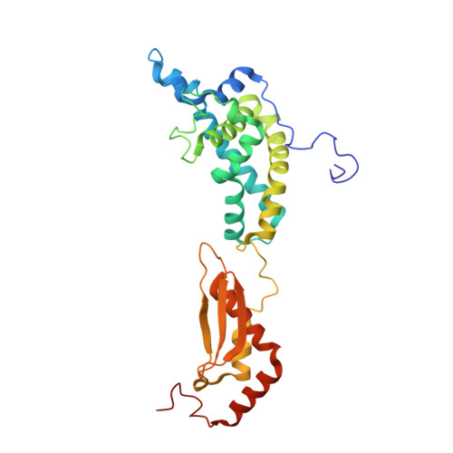
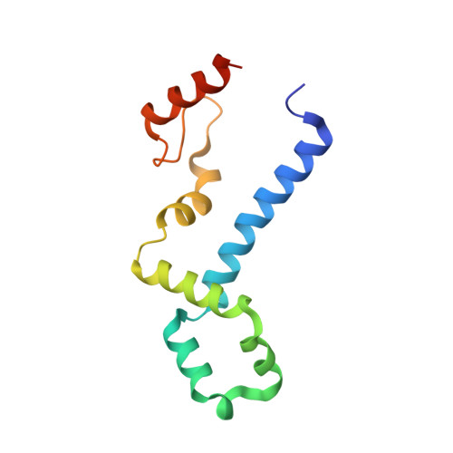
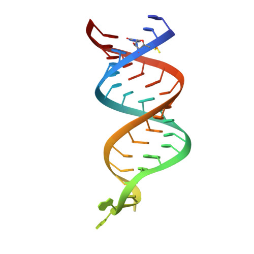
The Functional Cycle of Rnt1p: Five Consecutive Steps of Double-Stranded RNA Processing by a Eukaryotic RNase III.

Song, H.Fang, X.Jin, L.Shaw, G.X.Wang, Y.X.Ji, X.

(2017) Structure 25: 353-363

  • DOI: https://doi.org/10.1016/j.str.2016.12.013
  • Primary Citation Related Structures: 
    5T16

  • PubMed Abstract: 

    Double-stranded RNA (dsRNA)-specific RNase III proteins are required for RNA maturation and gene regulation. The mechanism of prokaryotic RNase IIIs has been well characterized, but how eukaryotic RNase IIIs (exemplified by Rnt1p, Drosha, and Dicer) work is less clear. Recently, we reported the crystal structure of Rnt1p in complex with RNA, revealing a double-ruler mechanism for substrate selection. Here, we present more structures of Rnt1p, either RNA free or RNA bound, featuring two major conformations of the enzyme. Using these structures with existing data, we describe the functional cycle of Rnt1p in five steps, selecting, loading, locking, cleavage, and releasing. We also describe atomic details of the two-Mg 2+ -ion catalytic mechanism that is applicable to all eukaryotic RNase III enzymes. Overall, our results indicate that substrate selection is achieved independent of cleavage, allowing the recognition of substrates with different structures while preserving the basic mechanism of cleavage.


  • Organizational Affiliation
    • Macromolecular Crystallography Laboratory, National Cancer Institute, Frederick, MD 21702, USA.

Macromolecule Content 

  • Total Structure Weight: 282.12 kDa 
  • Atom Count: 19,568 
  • Modeled Residue Count: 2,142 
  • Deposited Residue Count: 2,192 
  • Unique protein chains: 2
  • Unique nucleic acid chains: 1

Macromolecules


Find similar proteins by:|  3D Structure
Entity ID: 1
MoleculeChains  Sequence LengthOrganismDetailsImage
Ribonuclease 3
A, B, I, J
276Saccharomyces cerevisiae S288CMutation(s): 0 
Gene Names: RNT1YMR239CYM9408.01CYM9959.21
EC: 3.1.26.3
UniProt
Find proteins for Q02555 (Saccharomyces cerevisiae (strain ATCC 204508 / S288c))
Explore Q02555 
Go to UniProtKB:  Q02555
Entity Groups
Sequence Clusters30% Identity50% Identity70% Identity90% Identity95% Identity100% Identity
UniProt GroupQ02555
Sequence Annotations
Expand
Reference Sequence
Find similar proteins by:|  3D Structure
Entity ID: 2
MoleculeChains  Sequence LengthOrganismDetailsImage
Ribonuclease 3
C, D, E, F, K
C, D, E, F, K, L, M, N
119Saccharomyces cerevisiae S288CMutation(s): 0 
Gene Names: RNT1YMR239CYM9408.01CYM9959.21
EC: 3.1.26.3
UniProt
Find proteins for Q02555 (Saccharomyces cerevisiae (strain ATCC 204508 / S288c))
Explore Q02555 
Go to UniProtKB:  Q02555
Entity Groups
Sequence Clusters30% Identity50% Identity70% Identity90% Identity95% Identity100% Identity
UniProt GroupQ02555
Sequence Annotations
Expand
Reference Sequence
Find similar nucleic acids by:  (by identity cutoff) 
Entity ID: 3
MoleculeChains LengthOrganismImage
RNA substrate analog
G, H, O, P
34N/A
Sequence Annotations
Expand
Reference Sequence

Small Molecules

Ligands 1 Unique
IDChains Name / Formula / InChI Key2D Diagram3D Interactions
EDO

Query on EDO


Download:Ideal Coordinates CCD File
Q [auth A]
R [auth E]
S [auth F]
T [auth H]
U [auth I]
Q [auth A],
R [auth E],
S [auth F],
T [auth H],
U [auth I],
V [auth I],
W [auth I],
X [auth I],
Y [auth I],
Z [auth J]
1,2-ETHANEDIOL
C2 H6 O2
LYCAIKOWRPUZTN-UHFFFAOYSA-N

Experimental Data & Validation

Experimental Data

  • Method: X-RAY DIFFRACTION
  • Resolution: 2.78 Å
  • R-Value Free:  0.265 (Depositor), 0.266 (DCC) 
  • R-Value Work:  0.226 (Depositor), 0.227 (DCC) 
  • R-Value Observed: 0.226 (Depositor) 
Space Group: P 1 21 1
Unit Cell:
Length ( Å )Angle ( ˚ )
a = 62.037α = 90
b = 164.065β = 96.78
c = 176.855γ = 90
Software Package:
Software NamePurpose
PHENIXrefinement
HKL-3000data reduction
HKL-3000data scaling
PHASERphasing

Structure Validation

View Full Validation Report


Entry History 

Deposition Data

Revision History  (Full details and data files)

  • Version 1.0: 2017-02-08
    Type: Initial release
  • Version 1.1: 2017-02-15
    Changes: Database references
  • Version 1.2: 2023-08-30
    Changes: Data collection, Database references, Structure summary
  • Version 1.3: 2023-10-04
    Changes: Refinement description