5SYG | pdb_00005syg

Cryo-EM reconstruction of zampanolide-bound microtubule

  • Classification: STRUCTURAL PROTEIN
  • Organism(s): Sus scrofa
  • Mutation(s): No 

  • Deposited: 2016-08-11 Released: 2017-02-01 
  • Deposition Author(s): Kellogg, E.H., Hejab, N.M.A., Nogales, E.
  • Funding Organization(s): National Institutes of Health/National Institute of General Medical Sciences (NIH/NIGMS), Ministerio de Economia y Competitividad, Comunidad Autonoma de Madrid

Experimental Data Snapshot

  • Method: ELECTRON MICROSCOPY
  • Resolution: 3.50 Å
  • Aggregation State: HELICAL ARRAY 
  • Reconstruction Method: HELICAL 

wwPDB Validation 3D Report Full Report

Validation slider image for 5SYG

This is version 1.4 of the entry. See complete history

Literature

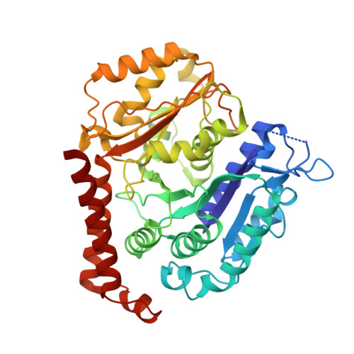
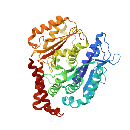
Insights into the Distinct Mechanisms of Action of Taxane and Non-Taxane Microtubule Stabilizers from Cryo-EM Structures.

Kellogg, E.H.Hejab, N.M.Howes, S.Northcote, P.Miller, J.H.Diaz, J.F.Downing, K.H.Nogales, E.

(2017) J Mol Biology 429: 633-646

  • DOI: https://doi.org/10.1016/j.jmb.2017.01.001
  • Primary Citation Related Structures: 
    5SYC, 5SYE, 5SYF, 5SYG

  • PubMed Abstract: 

    A number of microtubule (MT)-stabilizing agents (MSAs) have demonstrated or predicted potential as anticancer agents, but a detailed structural basis for their mechanism of action is still lacking. We have obtained high-resolution (3.9-4.2Å) cryo-electron microscopy (cryo-EM) reconstructions of MTs stabilized by the taxane-site binders Taxol and zampanolide, and by peloruside, which targets a distinct, non-taxoid pocket on β-tubulin. We find that each molecule has unique distinct structural effects on the MT lattice structure. Peloruside acts primarily at lateral contacts and has an effect on the "seam" of heterologous interactions, enforcing a conformation more similar to that of homologous (i.e., non-seam) contacts by which it regularizes the MT lattice. In contrast, binding of either Taxol or zampanolide induces MT heterogeneity. In doubly bound MTs, peloruside overrides the heterogeneity induced by Taxol binding. Our structural analysis illustrates distinct mechanisms of these drugs for stabilizing the MT lattice and is of relevance to the possible use of combinations of MSAs to regulate MT activity and improve therapeutic potential.


  • Organizational Affiliation
    • Molecular Biophysics and Integrated Bioimaging Division, Lawrence Berkeley National Laboratory, Berkeley, CA 94720, USA.

Macromolecule Content 

  • Total Structure Weight: 97.99 kDa 
  • Atom Count: 6,792 
  • Modeled Residue Count: 852 
  • Deposited Residue Count: 863 
  • Unique protein chains: 2

Macromolecules

Find similar proteins by:|  3D Structure
Entity ID: 1
MoleculeChains  Sequence LengthOrganismDetailsImage
Tubulin alpha chain437Sus scrofaMutation(s): 0 
EC: 3.6.5
UniProt
Find proteins for Q2XVP4 (Sus scrofa)
Explore Q2XVP4 
Go to UniProtKB:  Q2XVP4
Entity Groups
Sequence Clusters30% Identity50% Identity70% Identity90% Identity95% Identity100% Identity
UniProt GroupQ2XVP4
Sequence Annotations
Expand
Reference Sequence
Find similar proteins by:|  3D Structure
Entity ID: 2
MoleculeChains  Sequence LengthOrganismDetailsImage
Tubulin beta chain426Sus scrofaMutation(s): 0 
UniProt
Find proteins for P02554 (Sus scrofa)
Explore P02554 
Go to UniProtKB:  P02554
Entity Groups
Sequence Clusters30% Identity50% Identity70% Identity90% Identity95% Identity100% Identity
UniProt GroupP02554
Sequence Annotations
Expand
Reference Sequence

Small Molecules

Ligands 4 Unique
IDChains Name / Formula / InChI Key2D Diagram3D Interactions
GTP

Query on GTP


Download:Ideal Coordinates CCD File
C [auth A]GUANOSINE-5'-TRIPHOSPHATE
C10 H16 N5 O14 P3
XKMLYUALXHKNFT-UUOKFMHZSA-N
ZPN

Query on ZPN


Download:Ideal Coordinates CCD File
F [auth B](2Z,4E)-N-[(S)-[(1S,2E,5S,8E,10Z,17S)-3,11-dimethyl-19-methylidene-7,13-dioxo-6,21-dioxabicyclo[15.3.1]henicosa-2,8,10-trien-5-yl](hydroxy)methyl]hexa-2,4-dienamide
C29 H39 N O6
KNEMNZWQWFRUIB-KZVIXHMTSA-N
GDP

Query on GDP


Download:Ideal Coordinates CCD File
E [auth B]GUANOSINE-5'-DIPHOSPHATE
C10 H15 N5 O11 P2
QGWNDRXFNXRZMB-UUOKFMHZSA-N
MG

Query on MG


Download:Ideal Coordinates CCD File
D [auth A]MAGNESIUM ION
Mg
JLVVSXFLKOJNIY-UHFFFAOYSA-N

Experimental Data & Validation

Experimental Data

  • Method: ELECTRON MICROSCOPY
  • Resolution: 3.50 Å
  • Aggregation State: HELICAL ARRAY 
  • Reconstruction Method: HELICAL 
EM Software:
TaskSoftware PackageVersion
RECONSTRUCTIONFREALIGN9.09
MODEL REFINEMENTRosetta3.6
MODEL REFINEMENTREFMAC5.5

Structure Validation

View Full Validation Report


Entry History 

& Funding Information

Deposition Data


Funding OrganizationLocationGrant Number
National Institutes of Health/National Institute of General Medical Sciences (NIH/NIGMS)United StatesGM51487
Ministerio de Economia y CompetitividadSpainBIO2013-42984-R
Comunidad Autonoma de MadridSpainS2010/BMD-2457 BIPEDD2

Revision History  (Full details and data files)

  • Version 1.0: 2017-02-01
    Type: Initial release
  • Version 1.1: 2017-03-08
    Changes: Database references
  • Version 1.2: 2017-09-13
    Changes: Author supporting evidence, Data collection
  • Version 1.3: 2019-12-25
    Changes: Author supporting evidence
  • Version 1.4: 2024-03-06
    Changes: Data collection, Database references, Derived calculations