5SXS

Crystal structure of catalase-peroxidase KatG with isonicotinic acid hydrazide and AMP bound


Experimental Data Snapshot

  • Method: X-RAY DIFFRACTION
  • Resolution: 1.89 Å
  • R-Value Free: 0.193 
  • R-Value Work: 0.162 
  • R-Value Observed: 0.163 

Starting Model: experimental
View more details

wwPDB Validation   3D Report Full Report


Ligand Structure Quality Assessment 


This is version 1.3 of the entry. See complete history


Literature

Isonicotinic acid hydrazide conversion to Isonicotinyl-NAD by catalase-peroxidases.

Wiseman, B.Carpena, X.Feliz, M.Donald, L.J.Pons, M.Fita, I.Loewen, P.C.

(2010) J Biol Chem 285: 26662-26673

  • DOI: https://doi.org/10.1074/jbc.M110.139428
  • Primary Citation of Related Structures:  
    5SXQ, 5SXR, 5SXS, 5SXT, 5SXW, 5SXX

  • PubMed Abstract: 

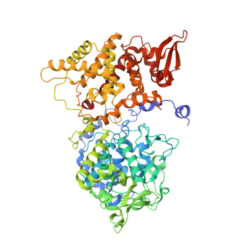
    Activation of the pro-drug isoniazid (INH) as an anti-tubercular drug in Mycobacterium tuberculosis involves its conversion to isonicotinyl-NAD, a reaction that requires the catalase-peroxidase KatG. This report shows that the reaction proceeds in the absence of KatG at a slow rate in a mixture of INH, NAD(+), Mn(2+), and O(2), and that the inclusion of KatG increases the rate by >7 times. Superoxide, generated by either Mn(2+)- or KatG-catalyzed reduction of O(2), is an essential intermediate in the reaction. Elimination of the peroxidatic process by mutation slows the rate of reaction by 60% revealing that the peroxidatic process enhances, but is not essential for isonicotinyl-NAD formation. The isonicotinyl-NAD(*+) radical is identified as a reaction intermediate, and its reduction by superoxide is proposed. Binding sites for INH and its co-substrate, NAD(+), are identified for the first time in crystal complexes of Burkholderia pseudomallei catalase-peroxidase with INH and NAD(+) grown by co-crystallization. The best defined INH binding sites were identified, one in each subunit, on the opposite side of the protein from the entrance to the heme cavity in a funnel-shaped channel. The NAD(+) binding site is approximately 20 A from the entrance to the heme cavity and involves interactions primarily with the AMP portion of the molecule in agreement with the NMR saturation transfer difference results.


  • Organizational Affiliation

    Department of Microbiology, University of Manitoba,Winnipeg, Manitoba, Canada.


Macromolecules
Find similar proteins by:  (by identity cutoff)  |  3D Structure
Entity ID: 1
MoleculeChains Sequence LengthOrganismDetailsImage
Catalase-peroxidase
A, B
728Burkholderia pseudomallei 1710bMutation(s): 0 
Gene Names: katGBURPS1710b_3366
EC: 1.11.1.21
UniProt
Find proteins for Q3JNW6 (Burkholderia pseudomallei (strain 1710b))
Explore Q3JNW6 
Go to UniProtKB:  Q3JNW6
Entity Groups  
Sequence Clusters30% Identity50% Identity70% Identity90% Identity95% Identity100% Identity
UniProt GroupQ3JNW6
Sequence Annotations
Expand
  • Reference Sequence
Small Molecules
Ligands 9 Unique
IDChains Name / Formula / InChI Key2D Diagram3D Interactions
HEM
Query on HEM

Download Ideal Coordinates CCD File 
C [auth A],
J [auth B]
PROTOPORPHYRIN IX CONTAINING FE
C34 H32 Fe N4 O4
KABFMIBPWCXCRK-RGGAHWMASA-L
AMP
Query on AMP

Download Ideal Coordinates CCD File 
R [auth B]ADENOSINE MONOPHOSPHATE
C10 H14 N5 O7 P
UDMBCSSLTHHNCD-KQYNXXCUSA-N
NIZ
Query on NIZ

Download Ideal Coordinates CCD File 
O [auth B]pyridine-4-carbohydrazide
C6 H7 N3 O
QRXWMOHMRWLFEY-UHFFFAOYSA-N
MPD
Query on MPD

Download Ideal Coordinates CCD File 
P [auth B](4S)-2-METHYL-2,4-PENTANEDIOL
C6 H14 O2
SVTBMSDMJJWYQN-YFKPBYRVSA-N
MRD
Query on MRD

Download Ideal Coordinates CCD File 
I [auth A](4R)-2-METHYLPENTANE-2,4-DIOL
C6 H14 O2
SVTBMSDMJJWYQN-RXMQYKEDSA-N
PO4
Query on PO4

Download Ideal Coordinates CCD File 
H [auth A],
Q [auth B]
PHOSPHATE ION
O4 P
NBIIXXVUZAFLBC-UHFFFAOYSA-K
CL
Query on CL

Download Ideal Coordinates CCD File 
E [auth A],
L [auth B]
CHLORIDE ION
Cl
VEXZGXHMUGYJMC-UHFFFAOYSA-M
OXY
Query on OXY

Download Ideal Coordinates CCD File 
F [auth A],
G [auth A],
M [auth B],
N [auth B]
OXYGEN MOLECULE
O2
MYMOFIZGZYHOMD-UHFFFAOYSA-N
NA
Query on NA

Download Ideal Coordinates CCD File 
D [auth A],
K [auth B]
SODIUM ION
Na
FKNQFGJONOIPTF-UHFFFAOYSA-N
Experimental Data & Validation

Experimental Data

  • Method: X-RAY DIFFRACTION
  • Resolution: 1.89 Å
  • R-Value Free: 0.193 
  • R-Value Work: 0.162 
  • R-Value Observed: 0.163 
  • Space Group: P 21 21 21
Unit Cell:
Length ( Å )Angle ( ˚ )
a = 100.436α = 90
b = 114.172β = 90
c = 174.232γ = 90
Software Package:
Software NamePurpose
REFMACrefinement
PDB_EXTRACTdata extraction
MOSFLMdata reduction
SCALAdata scaling
MOLREPphasing

Structure Validation

View Full Validation Report


Ligand Structure Quality Assessment 


Entry History & Funding Information

Deposition Data

  • Released Date: 2016-09-07 
  • Deposition Author(s): Loewen, P.C.
  • This entry supersedes: 3N3P

Funding OrganizationLocationGrant Number
Natural Sciences and Engineering Research Council (NSERC, Canada)CanadaD9600

Revision History  (Full details and data files)

  • Version 1.0: 2016-09-07
    Type: Initial release
  • Version 1.1: 2017-09-27
    Changes: Author supporting evidence, Derived calculations, Structure summary
  • Version 1.2: 2020-01-08
    Changes: Author supporting evidence
  • Version 1.3: 2023-10-04
    Changes: Data collection, Database references, Derived calculations, Refinement description