5SX4 | pdb_00005sx4

Crystal Structure of panitumumab in complex with epidermal growth factor receptor domain 3.


Experimental Data Snapshot

  • Method: X-RAY DIFFRACTION
  • Resolution: 2.80 Å
  • R-Value Free: 
    0.266 (Depositor), 0.267 (DCC) 
  • R-Value Work: 
    0.223 (Depositor), 0.223 (DCC) 
  • R-Value Observed: 
    0.226 (Depositor) 

Starting Model: experimental
View more details

wwPDB Validation 3D Report Full Report

Validation slider image for 5SX4

This is version 1.2 of the entry. See complete history

Literature

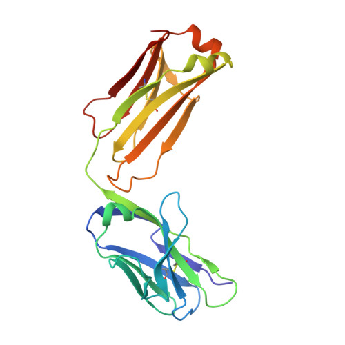
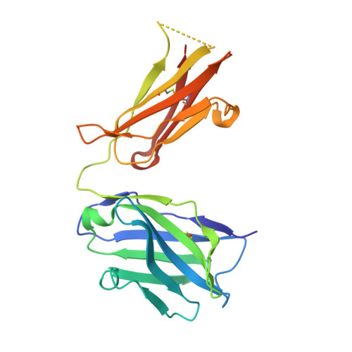
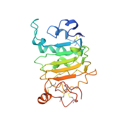
The Panitumumab EGFR Complex Reveals a Binding Mechanism That Overcomes Cetuximab Induced Resistance.

Sickmier, E.A.Kurzeja, R.J.Michelsen, K.Vazir, M.Yang, E.Tasker, A.S.

(2016) PLoS One 11: e0163366-e0163366

  • DOI: https://doi.org/10.1371/journal.pone.0163366
  • Primary Citation Related Structures: 
    5SX4, 5SX5

  • PubMed Abstract: 

    Panitumumab and cetuximab target the epidermal growth factor receptor for the treatment of metastatic colorectal cancer. These therapies provide a significant survival benefit to patients with metastatic colorectal cancer with wild-type RAS. A single point mutation in the ectodomain of EGFR (S468R) confers acquired or secondary resistance in cetuximab treated patients, which is not observed in panitumumab-treated patients. Structural and biophysical studies presented here show this mutation directly blocks cetuximab binding to EGFR domain III and describes a unique mechanism by which panitumumab uses a central cavity to accommodate this mutation.


  • Organizational Affiliation
    • Depatment of Therapeutic Discovery, Amgen Inc., 360 Binney Street, Cambridge, MA 02142, United States of America.

Macromolecule Content 

  • Total Structure Weight: 140.07 kDa 
  • Atom Count: 9,504 
  • Modeled Residue Count: 1,218 
  • Deposited Residue Count: 1,272 
  • Unique protein chains: 3

Macromolecules

Find similar proteins by:|  3D Structure
Entity ID: 1
MoleculeChains  Sequence LengthOrganismDetailsImage
Panitumumab Fab Light ChainA [auth I],
C [auth L]
214Homo sapiensMutation(s): 0 
Find similar proteins by:|  3D Structure
Entity ID: 2
MoleculeChains  Sequence LengthOrganismDetailsImage
Panitumumab Fab Heavy ChainB [auth J],
D [auth H]
221Homo sapiensMutation(s): 0 
Find similar proteins by:|  3D Structure
Entity ID: 3
MoleculeChains  Sequence LengthOrganismDetailsImage
Epidermal growth factor receptorE [auth M],
F [auth N]
201Homo sapiensMutation(s): 0 
Gene Names: EGFRERBBERBB1HER1
EC: 2.7.10.1
UniProt & NIH Common Fund Data Resources
Find proteins for P00533 (Homo sapiens)
Explore P00533 
Go to UniProtKB:  P00533
PHAROS:  P00533
GTEx:  ENSG00000146648 
Entity Groups
Sequence Clusters30% Identity50% Identity70% Identity90% Identity95% Identity100% Identity
UniProt GroupP00533
Sequence Annotations
Expand
Reference Sequence

Small Molecules

Ligands 4 Unique
IDChains Name / Formula / InChI Key2D Diagram3D Interactions
1PE

Query on 1PE


Download:Ideal Coordinates CCD File
K [auth J],
P [auth H]
PENTAETHYLENE GLYCOL
C10 H22 O6
JLFNLZLINWHATN-UHFFFAOYSA-N
SO4

Query on SO4


Download:Ideal Coordinates CCD File
G [auth I]
H [auth I]
I
L
M [auth L]
G [auth I],
H [auth I],
I,
L,
M [auth L],
N [auth L],
O [auth L],
R [auth N]
SULFATE ION
O4 S
QAOWNCQODCNURD-UHFFFAOYSA-L
GOL

Query on GOL


Download:Ideal Coordinates CCD File
J [auth I]GLYCEROL
C3 H8 O3
PEDCQBHIVMGVHV-UHFFFAOYSA-N
EDO

Query on EDO


Download:Ideal Coordinates CCD File
Q [auth H]1,2-ETHANEDIOL
C2 H6 O2
LYCAIKOWRPUZTN-UHFFFAOYSA-N

Experimental Data & Validation

Experimental Data

  • Method: X-RAY DIFFRACTION
  • Resolution: 2.80 Å
  • R-Value Free:  0.266 (Depositor), 0.267 (DCC) 
  • R-Value Work:  0.223 (Depositor), 0.223 (DCC) 
  • R-Value Observed: 0.226 (Depositor) 
Space Group: P 21 21 21
Unit Cell:
Length ( Å )Angle ( ˚ )
a = 65.273α = 90
b = 113.184β = 90
c = 232.443γ = 90
Software Package:
Software NamePurpose
SCALEPACKdata scaling
PHENIXrefinement
PDB_EXTRACTdata extraction
HKL-2000data reduction
PHASERphasing

Structure Validation

View Full Validation Report


Entry History 

Deposition Data

Revision History  (Full details and data files)

  • Version 1.0: 2016-10-05
    Type: Initial release
  • Version 1.1: 2023-10-04
    Changes: Data collection, Database references, Derived calculations, Refinement description
  • Version 1.2: 2024-10-16
    Changes: Structure summary