


Crystal structure of human Cathepsin-S with bound ligand
Bembenek, S.D., Ameriks, M.K., Mirzadegan, T., Yang, H., Shao, C., Burley, S.K.To be published.
Experimental Data Snapshot
Entity ID: 1 | |||||
|---|---|---|---|---|---|
| Molecule | Chains | Sequence Length | Organism | Details | Image |
| Cathepsin S | 223 | Homo sapiens | Mutation(s): 1 Gene Names: CTSS EC: 3.4.22.27 | 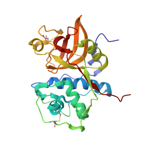 | |
UniProt & NIH Common Fund Data Resources | |||||
PHAROS: P25774 GTEx: ENSG00000163131 | |||||
Entity Groups | |||||
| Sequence Clusters | 30% Identity50% Identity70% Identity90% Identity95% Identity100% Identity | ||||
| UniProt Group | P25774 | ||||
Sequence AnnotationsExpand | |||||
Reference Sequence | |||||
| Ligands 2 Unique | |||||
|---|---|---|---|---|---|
| ID | Chains | Name / Formula / InChI Key | 2D Diagram | 3D Interactions | |
| BHJ Download:Ideal Coordinates CCD File | B [auth A] | {(3S)-7-[(2-chloro-5-{5-(methylsulfonyl)-1-[3-(morpholin-4-yl)propyl]-4,5,6,7-tetrahydro-1H-pyrazolo[4,3-c]pyridin-3-yl}phenyl)ethynyl]-1,2,3,4-tetrahydroisoquinolin-3-yl}(piperidin-1-yl)methanone C37 H45 Cl N6 O4 S BIYANWMTXPESQI-UMSFTDKQSA-N |  | ||
| SO4 Download:Ideal Coordinates CCD File | C [auth A] | SULFATE ION O4 S QAOWNCQODCNURD-UHFFFAOYSA-L |  | ||
| Length ( Å ) | Angle ( ˚ ) |
|---|---|
| a = 76.952 | α = 90 |
| b = 73.329 | β = 103.97 |
| c = 44.594 | γ = 90 |
| Software Name | Purpose |
|---|---|
| PHENIX | refinement |
| PDB_EXTRACT | data extraction |
| MOSFLM | data reduction |
| SCALA | data scaling |


