5QBV | pdb_00005qbv

Crystal structure of human Cathepsin-S with bound ligand


Experimental Data Snapshot

  • Method: X-RAY DIFFRACTION
  • Resolution: 1.80 Å
  • R-Value Free: 
    0.172 (Depositor), 0.173 (DCC) 
  • R-Value Work: 
    0.147 (Depositor), 0.148 (DCC) 
  • R-Value Observed: 
    0.148 (Depositor) 

wwPDB Validation 3D Report Full Report

Validation slider image for 5QBV

Ligand Structure Quality Assessment 


This is version 1.4 of the entry. See complete history

Literature

Diazinones as P2 replacements for pyrazole-based cathepsin S inhibitors

Ameriks, M.K.Bembenek, S.D.Burdett, M.T.Choong, I.C.Edwards, J.P.Gebauer, D.Gu, Y.Karlsson, L.Purkey, H.E.Staker, B.L.Sun, S.Thurmond, R.L.Zhu, J.

(2010) Bioorg Med Chem Lett 20: 4060-4064

  • DOI: https://doi.org/10.1016/j.bmcl.2010.05.086
  • Primary Citation Related Structures: 
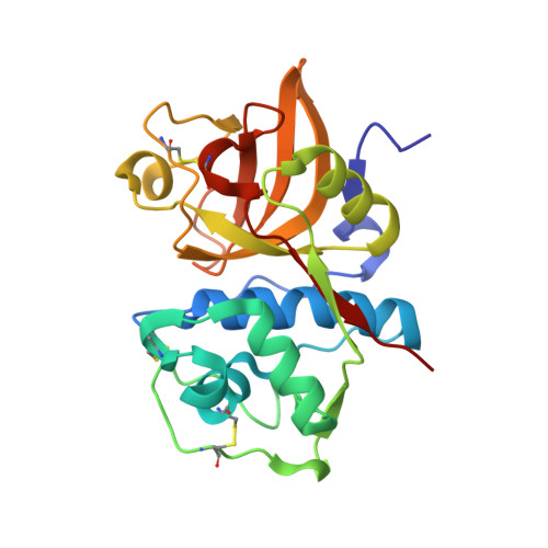
    5QBV, 5QBY

  • PubMed Abstract: 

    A pyridazin-4-one fragment 4 (hCatS IC(50)=170 microM) discovered through Tethering was modeled into cathepsin S and predicted to overlap in S2 with the tetrahydropyridinepyrazole core of a previously disclosed series of CatS inhibitors. This fragment served as a template to design pyridazin-3-one 12 (hCatS IC(50)=430 nM), which also incorporates P3 and P5 binding elements. A crystal structure of 12 bound to Cys25Ser CatS led to the synthesis of the potent diazinone isomers 22 (hCatS IC(50)=60 nM) and 27 (hCatS IC(50)=40 nM).


  • Organizational Affiliation
    • Johnson & Johnson Pharmaceutical Research & Development, L.L.C., 3210 Merryfield Row, San Diego, CA 92121, USA. mameriks@its.jnj.com

Macromolecule Content 

  • Total Structure Weight: 50.32 kDa 
  • Atom Count: 4,098 
  • Modeled Residue Count: 439 
  • Deposited Residue Count: 446 
  • Unique protein chains: 1

Macromolecules

Find similar proteins by:|  3D Structure
Entity ID: 1
MoleculeChains  Sequence LengthOrganismDetailsImage
Cathepsin S
A, B
223Homo sapiensMutation(s): 1 
Gene Names: CTSS
EC: 3.4.22.27
UniProt & NIH Common Fund Data Resources
Find proteins for P25774 (Homo sapiens)
Explore P25774 
Go to UniProtKB:  P25774
PHAROS:  P25774
GTEx:  ENSG00000163131 
Entity Groups
Sequence Clusters30% Identity50% Identity70% Identity90% Identity95% Identity100% Identity
UniProt GroupP25774
Sequence Annotations
Expand
Reference Sequence

Small Molecules

Ligands 1 Unique
IDChains Name / Formula / InChI Key2D Diagram3D Interactions
N2D

Query on N2D


Download:Ideal Coordinates CCD File
C [auth A],
D [auth B]
N-[2-chloro-5-(1-{3-[4-(6-chloro-3-methyl-2-oxo-2,3-dihydro-1H-benzimidazol-1-yl)piperidin-1-yl]propyl}-6-oxo-1,6-dihydropyridazin-3-yl)benzyl]benzamide
C34 H34 Cl2 N6 O3
ZJXORVFCSQEKCZ-UHFFFAOYSA-N
Binding Affinity Annotations 
IDSourceBinding Affinity
N2D BindingDB:  5QBV IC50: 430 (nM) from 1 assay(s)

Experimental Data & Validation

Experimental Data

  • Method: X-RAY DIFFRACTION
  • Resolution: 1.80 Å
  • R-Value Free:  0.172 (Depositor), 0.173 (DCC) 
  • R-Value Work:  0.147 (Depositor), 0.148 (DCC) 
  • R-Value Observed: 0.148 (Depositor) 
Space Group: C 1 2 1
Unit Cell:
Length ( Å )Angle ( ˚ )
a = 172.391α = 90
b = 34.311β = 125.27
c = 104.021γ = 90
Software Package:
Software NamePurpose
PHENIXrefinement
PDB_EXTRACTdata extraction
MOSFLMdata reduction
SCALAdata scaling

Structure Validation

View Full Validation Report


Ligand Structure Quality Assessment 


Entry History 

Deposition Data

Revision History  (Full details and data files)

  • Version 1.0: 2017-12-20
    Type: Initial release
  • Version 1.1: 2018-02-21
    Changes: Structure summary
  • Version 1.2: 2021-02-10
    Changes: Structure summary
  • Version 1.3: 2021-11-17
    Changes: Database references, Structure summary
  • Version 1.4: 2024-10-16
    Changes: Data collection, Refinement description, Structure summary