5OWY | pdb_00005owy

Glycogen Phosphorylase in complex with KS252


Experimental Data Snapshot

  • Method: X-RAY DIFFRACTION
  • Resolution: 1.90 Å
  • R-Value Free: 
    0.163 (Depositor), 0.170 (DCC) 
  • R-Value Work: 
    0.135 (Depositor), 0.150 (DCC) 
  • R-Value Observed: 
    0.137 (Depositor) 

wwPDB Validation   3D Report Full Report


Ligand Structure Quality Assessment 

Created with Raphaël 2.3.0Worse 01 BetterLigand structure goodness of fit to experimental dataBest fitted B0WClick on this verticalbar to view detailsBest fitted IMPClick on this verticalbar to view detailsBest fitted PLPClick on this verticalbar to view details

This is version 1.1 of the entry. See complete history


Literature

Probing the beta-pocket of the active site of human liver glycogen phosphorylase with 3-(C-beta-d-glucopyranosyl)-5-(4-substituted-phenyl)-1, 2, 4-triazole inhibitors.

Kyriakis, E.Solovou, T.G.A.Kun, S.Czifrak, K.Szocs, B.Juhasz, L.Bokor, E.Stravodimos, G.A.Kantsadi, A.L.Chatzileontiadou, D.S.M.Skamnaki, V.T.Somsak, L.Leonidas, D.D.

(2018) Bioorg Chem 77: 485-493

  • DOI: https://doi.org/10.1016/j.bioorg.2018.02.008
  • Primary Citation of Related Structures:  
    5OWY, 5OWZ, 5OX0, 5OX1, 5OX3, 5OX4

  • PubMed Abstract: 

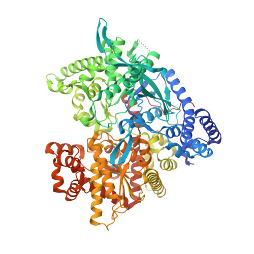
    Human liver glycogen phosphorylase (hlGP), a key enzyme in glycogen metabolism, is a valid pharmaceutical target for the development of new anti-hyperglycaemic agents for type 2 diabetes. Inhibitor discovery studies have focused on the active site and in particular on glucopyranose based compounds with a β-1 substituent long enough to exploit interactions with a cavity adjacent to the active site, termed the β-pocket. Recently, C-β-d-glucopyranosyl imidazoles and 1, 2, 4-triazoles proved to be the best known glucose derived inhibitors of hlGP. Here we probe the β-pocket by studying the inhibitory effect of six different groups at the para position of 3-(β-d-glucopyranosyl phenyl)-5-phenyl-, 1, 2, 4-triazoles in hlGP by kinetics and X-ray crystallography. The most bioactive compound was the one with an amine substituent to show a K i value of 0.43 μM. Structural studies have revealed the physicochemical diversity of the β-pocket providing information for future rational inhibitor design studies.


  • Organizational Affiliation

    Department of Biochemistry and Biotechnology, University of Thessaly, Biopolis, 41500 Larissa, Greece.


Macromolecules
Find similar proteins by:  (by identity cutoff)  |  3D Structure
Entity ID: 1
MoleculeChains Sequence LengthOrganismDetailsImage
Glycogen phosphorylase, muscle form843Oryctolagus cuniculusMutation(s): 0 
EC: 2.4.1.1
UniProt
Find proteins for P00489 (Oryctolagus cuniculus)
Explore P00489 
Go to UniProtKB:  P00489
Entity Groups  
Sequence Clusters30% Identity50% Identity70% Identity90% Identity95% Identity100% Identity
UniProt GroupP00489
Sequence Annotations
Expand
  • Reference Sequence
Small Molecules
Ligands 3 Unique
IDChains Name / Formula / InChI Key2D Diagram3D Interactions
B0W
Query on B0W

Download Ideal Coordinates CCD File 
C [auth A]4-[5-[(2~{S},3~{R},4~{R},5~{S},6~{R})-6-(hydroxymethyl)-3,4,5-tris(oxidanyl)oxan-2-yl]-4~{H}-1,2,4-triazol-3-yl]benzoic acid
C15 H17 N3 O7
UYSDRGPJXJKBOO-RMPHRYRLSA-N
IMP
Query on IMP

Download Ideal Coordinates CCD File 
D [auth A]INOSINIC ACID
C10 H13 N4 O8 P
GRSZFWQUAKGDAV-KQYNXXCUSA-N
PLP
Query on PLP

Download Ideal Coordinates CCD File 
B [auth A]PYRIDOXAL-5'-PHOSPHATE
C8 H10 N O6 P
NGVDGCNFYWLIFO-UHFFFAOYSA-N
Experimental Data & Validation

Experimental Data

  • Method: X-RAY DIFFRACTION
  • Resolution: 1.90 Å
  • R-Value Free:  0.163 (Depositor), 0.170 (DCC) 
  • R-Value Work:  0.135 (Depositor), 0.150 (DCC) 
  • R-Value Observed: 0.137 (Depositor) 
Space Group: P 43 21 2
Unit Cell:
Length ( Å )Angle ( ˚ )
a = 128.46α = 90
b = 128.46β = 90
c = 116.19γ = 90
Software Package:
Software NamePurpose
REFMACrefinement
MOSFLMdata reduction
SCALAdata scaling
REFMACphasing

Structure Validation

View Full Validation Report


Ligand Structure Quality Assessment 

Created with Raphaël 2.3.0Worse 01 BetterLigand structure goodness of fit to experimental dataBest fitted B0WClick on this verticalbar to view detailsBest fitted IMPClick on this verticalbar to view detailsBest fitted PLPClick on this verticalbar to view details

Entry History 

Deposition Data

Revision History  (Full details and data files)

  • Version 1.0: 2018-02-28
    Type: Initial release
  • Version 1.1: 2025-04-09
    Changes: Data collection, Database references, Structure summary