5OWF | pdb_00005owf

Structure of a LAO-binding protein mutant with glutamine


Experimental Data Snapshot

  • Method: X-RAY DIFFRACTION
  • Resolution: 1.91 Å
  • R-Value Free: 
    0.231 (Depositor), 0.232 (DCC) 
  • R-Value Work: 
    0.190 (Depositor), 0.190 (DCC) 
  • R-Value Observed: 
    0.192 (Depositor) 

Starting Model: experimental
View more details

wwPDB Validation 3D Report Full Report

Validation slider image for 5OWF

Ligand Structure Quality Assessment 


This is version 1.5 of the entry. See complete history

Literature

Redesign of LAOBP to bind novel l-amino acid ligands.

Banda-Vazquez, J.Shanmugaratnam, S.Rodriguez-Sotres, R.Torres-Larios, A.Hocker, B.Sosa-Peinado, A.

(2018) Protein Sci 27: 957-968

  • DOI: https://doi.org/10.1002/pro.3403
  • Primary Citation Related Structures: 
    5OWF

  • PubMed Abstract: 

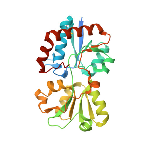
    Computational protein design is still a challenge for advancing structure-function relationships. While recent advances in this field are promising, more information for genuine predictions is needed. Here, we discuss different approaches applied to install novel glutamine (Gln) binding into the Lysine/Arginine/Ornithine binding protein (LAOBP) from Salmonella typhimurium. We studied the ligand binding behavior of two mutants: a binding pocket grafting design based on a structural superposition of LAOBP to the Gln binding protein QBP from Escherichia coli and a design based on statistical coupled positions. The latter showed the ability to bind Gln even though the protein was not very stable. Comparison of both approaches highlighted a nonconservative shared point mutation between LAOBP_graft and LAOBP_sca. This context dependent L117K mutation in LAOBP turned out to be sufficient for introducing Gln binding, as confirmed by different experimental techniques. Moreover, the crystal structure of LAOBP_L117K in complex with its ligand is reported.


  • Organizational Affiliation
    • Use National Autonomous University of Mexico, Mexico.

Macromolecule Content 

  • Total Structure Weight: 29.93 kDa 
  • Atom Count: 2,073 
  • Modeled Residue Count: 235 
  • Deposited Residue Count: 259 
  • Unique protein chains: 1

Macromolecules

Find similar proteins by:|  3D Structure
Entity ID: 1
MoleculeChains  Sequence LengthOrganismDetailsImage
Lysine/arginine/ornithine-binding periplasmic protein259Salmonella enterica subsp. enterica serovar Typhimurium str. LT2Mutation(s): 1 
Gene Names: argTSTM2355
UniProt
Find proteins for P02911 (Salmonella typhimurium (strain LT2 / SGSC1412 / ATCC 700720))
Explore P02911 
Go to UniProtKB:  P02911
Entity Groups
Sequence Clusters30% Identity50% Identity70% Identity90% Identity95% Identity100% Identity
UniProt GroupP02911
Sequence Annotations
Expand
Reference Sequence

Small Molecules

Ligands 4 Unique
IDChains Name / Formula / InChI Key2D Diagram3D Interactions
GLN
(Subject of Investigation/LOI)

Query on GLN


Download:Ideal Coordinates CCD File
U [auth A]GLUTAMINE
C5 H10 N2 O3
ZDXPYRJPNDTMRX-VKHMYHEASA-N
PEG

Query on PEG


Download:Ideal Coordinates CCD File
T [auth A]DI(HYDROXYETHYL)ETHER
C4 H10 O3
MTHSVFCYNBDYFN-UHFFFAOYSA-N
GOL

Query on GOL


Download:Ideal Coordinates CCD File
V [auth A],
W [auth A]
GLYCEROL
C3 H8 O3
PEDCQBHIVMGVHV-UHFFFAOYSA-N
EDO

Query on EDO


Download:Ideal Coordinates CCD File
B [auth A]
C [auth A]
D [auth A]
E [auth A]
F [auth A]
B [auth A],
C [auth A],
D [auth A],
E [auth A],
F [auth A],
G [auth A],
H [auth A],
I [auth A],
J [auth A],
K [auth A],
L [auth A],
M [auth A],
N [auth A],
O [auth A],
P [auth A],
Q [auth A],
R [auth A],
S [auth A]
1,2-ETHANEDIOL
C2 H6 O2
LYCAIKOWRPUZTN-UHFFFAOYSA-N

Experimental Data & Validation

Experimental Data

  • Method: X-RAY DIFFRACTION
  • Resolution: 1.91 Å
  • R-Value Free:  0.231 (Depositor), 0.232 (DCC) 
  • R-Value Work:  0.190 (Depositor), 0.190 (DCC) 
  • R-Value Observed: 0.192 (Depositor) 
Space Group: P 21 21 21
Unit Cell:
Length ( Å )Angle ( ˚ )
a = 36.972α = 90
b = 58.73β = 90
c = 115.312γ = 90
Software Package:
Software NamePurpose
XDSdata reduction
PHENIXrefinement
XDSdata scaling
PHENIXphasing

Structure Validation

View Full Validation Report


Ligand Structure Quality Assessment 


Entry History 

& Funding Information

Deposition Data


Funding OrganizationLocationGrant Number
German Research FoundationGermanyHO 4022/2-3
CONACyTMexico167838
PapiitMexicoIN214512-3

Revision History  (Full details and data files)

  • Version 1.0: 2018-03-21
    Type: Initial release
  • Version 1.1: 2018-04-18
    Changes: Data collection, Database references, Derived calculations, Source and taxonomy, Structure summary
  • Version 1.2: 2018-04-25
    Changes: Data collection, Derived calculations, Structure summary
  • Version 1.3: 2018-05-02
    Changes: Data collection, Database references
  • Version 1.4: 2024-01-17
    Changes: Data collection, Database references, Refinement description
  • Version 1.5: 2024-11-06
    Changes: Structure summary