5OV9 | pdb_00005ov9

Crystal structure of Acetylcholinesterase in complex with Crystal Violet


Experimental Data Snapshot

  • Method: X-RAY DIFFRACTION
  • Resolution: 2.40 Å
  • R-Value Free: 
    0.195 (Depositor), 0.166 (DCC) 
  • R-Value Work: 
    0.159 (Depositor), 0.173 (DCC) 
  • R-Value Observed: 
    0.159 (Depositor) 

Starting Model: experimental
View more details

wwPDB Validation 3D Report Full Report

Validation slider image for 5OV9

Ligand Structure Quality Assessment 


This is version 1.5 of the entry. See complete history

Literature

An Unusual Dimeric Inhibitor of Acetylcholinesterase: Cooperative Binding of Crystal Violet.

Allgardsson, A.David Andersson, C.Akfur, C.Worek, F.Linusson, A.Ekstrom, F.

(2017) Molecules 22

  • DOI: https://doi.org/10.3390/molecules22091433
  • Primary Citation Related Structures: 
    5OV9

  • PubMed Abstract: 

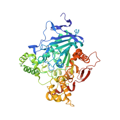
    Acetylcholinesterase (AChE) is an essential enzyme that terminates cholinergic transmission by a rapid hydrolysis of the neurotransmitter acetylcholine. AChE is an important target for treatment of various cholinergic deficiencies, including Alzheimer's disease and myasthenia gravis. In a previous high throughput screening campaign, we identified the dye crystal violet (CV) as an inhibitor of AChE. Herein, we show that CV displays a significant cooperativity for binding to AChE, and the molecular basis for this observation has been investigated by X-ray crystallography. Two monomers of CV bind to residues at the entrance of the active site gorge of the enzyme. Notably, the two CV molecules have extensive intermolecular contacts with each other and with AChE. Computational analyses show that the observed CV dimer is not stable in solution, suggesting the sequential binding of two monomers. Guided by the structural analysis, we designed a set of single site substitutions, and investigated their effect on the binding of CV. Only moderate effects on the binding and the cooperativity were observed, suggesting a robustness in the interaction between CV and AChE. Taken together, we propose that the dimeric cooperative binding is due to a rare combination of chemical and structural properties of both CV and the AChE molecule itself.


  • Organizational Affiliation
    • Swedish Defence Research Agency, SE-90281 Umeå, Sweden. anders.allgardsson@foi.se.

Macromolecule Content 

  • Total Structure Weight: 123.9 kDa 
  • Atom Count: 9,130 
  • Modeled Residue Count: 1,065 
  • Deposited Residue Count: 1,096 
  • Unique protein chains: 1

Macromolecules

Find similar proteins by:|  3D Structure
Entity ID: 1
MoleculeChains  Sequence LengthOrganismDetailsImage
Acetylcholinesterase
A, B
548Mus musculusMutation(s): 0 
Gene Names: Ache
EC: 3.1.1.7
UniProt & NIH Common Fund Data Resources
Find proteins for P21836 (Mus musculus)
Explore P21836 
Go to UniProtKB:  P21836
IMPC:  MGI:87876
Entity Groups
Sequence Clusters30% Identity50% Identity70% Identity90% Identity95% Identity100% Identity
UniProt GroupP21836
Glycosylation
Glycosylation Sites: 1Go to GlyGen: P21836-1
Sequence Annotations
Expand
Reference Sequence

Small Molecules

Ligands 9 Unique
IDChains Name / Formula / InChI Key2D Diagram3D Interactions
CVI

Query on CVI


Download:Ideal Coordinates CCD File
J [auth A],
K [auth A],
M [auth B],
N [auth B]
CRYSTAL VIOLET
C25 H30 N3
LGLFFNDHMLKUMI-UHFFFAOYSA-N
PE4

Query on PE4


Download:Ideal Coordinates CCD File
R [auth B]2-{2-[2-(2-{2-[2-(2-ETHOXY-ETHOXY)-ETHOXY]-ETHOXY}-ETHOXY)-ETHOXY]-ETHOXY}-ETHANOL
C16 H34 O8
PJWQOENWHPEPKI-UHFFFAOYSA-N
NAG

Query on NAG


Download:Ideal Coordinates CCD File
C [auth A],
L [auth B]
2-acetamido-2-deoxy-beta-D-glucopyranose
C8 H15 N O6
OVRNDRQMDRJTHS-FMDGEEDCSA-N
PGE

Query on PGE


Download:Ideal Coordinates CCD File
P [auth B]TRIETHYLENE GLYCOL
C6 H14 O4
ZIBGPFATKBEMQZ-UHFFFAOYSA-N
AE3

Query on AE3


Download:Ideal Coordinates CCD File
Q [auth B]2-(2-ETHOXYETHOXY)ETHANOL
C6 H14 O3
XXJWXESWEXIICW-UHFFFAOYSA-N
PG0

Query on PG0


Download:Ideal Coordinates CCD File
D [auth A],
E [auth A]
2-(2-METHOXYETHOXY)ETHANOL
C5 H12 O3
SBASXUCJHJRPEV-UHFFFAOYSA-N
PEG

Query on PEG


Download:Ideal Coordinates CCD File
O [auth B]DI(HYDROXYETHYL)ETHER
C4 H10 O3
MTHSVFCYNBDYFN-UHFFFAOYSA-N
ETX

Query on ETX


Download:Ideal Coordinates CCD File
F [auth A],
G [auth A],
S [auth B]
2-ETHOXYETHANOL
C4 H10 O2
ZNQVEEAIQZEUHB-UHFFFAOYSA-N
EDO

Query on EDO


Download:Ideal Coordinates CCD File
H [auth A],
I [auth A],
T [auth B],
U [auth B]
1,2-ETHANEDIOL
C2 H6 O2
LYCAIKOWRPUZTN-UHFFFAOYSA-N

Experimental Data & Validation

Experimental Data

  • Method: X-RAY DIFFRACTION
  • Resolution: 2.40 Å
  • R-Value Free:  0.195 (Depositor), 0.166 (DCC) 
  • R-Value Work:  0.159 (Depositor), 0.173 (DCC) 
  • R-Value Observed: 0.159 (Depositor) 
Space Group: P 21 21 21
Unit Cell:
Length ( Å )Angle ( ˚ )
a = 79.63α = 90
b = 113.89β = 90
c = 226.67γ = 90
Software Package:
Software NamePurpose
SCALAdata scaling
PHENIXrefinement
PDB_EXTRACTdata extraction
XDSdata reduction
REFMACphasing

Structure Validation

View Full Validation Report


Ligand Structure Quality Assessment 


Entry History 

& Funding Information

Deposition Data


Funding OrganizationLocationGrant Number
Swedish Research CouncilSweden2014-4675
Swedish Ministry of DefenceSweden--

Revision History  (Full details and data files)

  • Version 1.0: 2017-09-06
    Type: Initial release
  • Version 1.1: 2017-09-20
    Changes: Database references
  • Version 1.2: 2020-07-29
    Type: Remediation
    Reason: Carbohydrate remediation
    Changes: Data collection, Derived calculations, Structure summary
  • Version 1.3: 2021-05-12
    Changes: Derived calculations, Structure summary
  • Version 1.4: 2024-01-17
    Changes: Advisory, Data collection, Database references, Refinement description
  • Version 1.5: 2024-11-20
    Changes: Structure summary