Structure and activity of ChiX: a peptidoglycan hydrolase required for chitinase secretion by Serratia marcescens.
Owen, R.A., Fyfe, P.K., Lodge, A., Biboy, J., Vollmer, W., Hunter, W.N., Sargent, F.(2018) Biochem J 475: 415-428
- PubMed: 29229757 Search on PubMedSearch on PubMed Central
- DOI: https://doi.org/10.1042/BCJ20170633
- Primary Citation Related Structures:
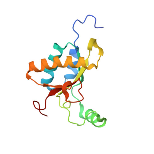
5OPZ, 5OQ1 - PubMed Abstract:
The Gram-negative bacterium Serratia marcescens secretes many proteins that are involved in extracellular chitin degradation. This so-called chitinolytic machinery includes three types of chitinase enzymes and a lytic polysaccharide monooxygenase. An operon has been identified in S. marcescens , chiWXYZ , that is thought to be involved in the secretion of the chitinolytic machinery. Genetic evidence points to the ChiX protein being a key player in the secretion mechanism, since deletion of the chiX gene in S. marcescens led to a mutant strain blocked for secretion of all members of the chitinolytic machinery. In this work, a detailed structural and biochemical characterisation of ChiX is presented. The high-resolution crystal structure of ChiX reveals the protein to be a member of the LAS family of peptidases. ChiX is shown to be a zinc-containing metalloenzyme, and in vitro assays demonstrate that ChiX is an l-Ala d-Glu endopeptidase that cleaves the cross-links in bacterial peptidoglycan. This catalytic activity is shown to be intimately linked with the secretion of the chitinolytic machinery, since substitution of the ChiX Asp-120 residue results in a variant protein that is both unable to digest peptidoglycan and cannot rescue the phenoytype of a chiX mutant strain.
- School of Life Sciences, University of Dundee, Dundee DD1 5EH, U.K.
Organizational Affiliation: