5OO7 | pdb_00005oo7

The ENTH domain from epsin-2 in complex with phosphatidylinositol 4,5-bisphosphate (PIP2)


Experimental Data Snapshot

  • Method: X-RAY DIFFRACTION
  • Resolution: 1.84 Å
  • R-Value Free: 
    0.252 (Depositor), 0.254 (DCC) 
  • R-Value Work: 
    0.204 (Depositor), 0.211 (DCC) 
  • R-Value Observed: 
    0.206 (Depositor) 

Starting Model: experimental
View more details

wwPDB Validation 3D Report Full Report

Validation slider image for 5OO7

This is version 1.2 of the entry. See complete history

Literature

Epsin and Sla2 form assemblies through phospholipid interfaces.

Garcia-Alai, M.M.Heidemann, J.Skruzny, M.Gieras, A.Mertens, H.D.T.Svergun, D.I.Kaksonen, M.Uetrecht, C.Meijers, R.

(2018) Nat Commun 9: 328-328

  • DOI: https://doi.org/10.1038/s41467-017-02443-x
  • Primary Citation Related Structures: 
    5ON7, 5ONF, 5OO7, 6ENR

  • PubMed Abstract: 

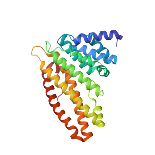
    In clathrin-mediated endocytosis, adapter proteins assemble together with clathrin through interactions with specific lipids on the plasma membrane. However, the precise mechanism of adapter protein assembly at the cell membrane is still unknown. Here, we show that the membrane-proximal domains ENTH of epsin and ANTH of Sla2 form complexes through phosphatidylinositol 4,5-bisphosphate (PIP2) lipid interfaces. Native mass spectrometry reveals how ENTH and ANTH domains form assemblies by sharing PIP2 molecules. Furthermore, crystal structures of epsin Ent2 ENTH domain from S. cerevisiae in complex with PIP2 and Sla2 ANTH domain from C. thermophilum illustrate how allosteric phospholipid binding occurs. A comparison with human ENTH and ANTH domains reveal only the human ENTH domain can form a stable hexameric core in presence of PIP2, which could explain functional differences between fungal and human epsins. We propose a general phospholipid-driven multifaceted assembly mechanism tolerating different adapter protein compositions to induce endocytosis.


  • Organizational Affiliation
    • European Molecular Biology Laboratory (EMBL), Hamburg Outstation, Notkestrasse 85, 22607, Hamburg, Germany.

Macromolecule Content 

  • Total Structure Weight: 59.44 kDa 
  • Atom Count: 4,615 
  • Modeled Residue Count: 513 
  • Deposited Residue Count: 514 
  • Unique protein chains: 1

Macromolecules

Find similar proteins by:|  3D Structure
Entity ID: 1
MoleculeChains  Sequence LengthOrganismDetailsImage
SLA2
A, B
257Thermochaetoides thermophila DSM 1495Mutation(s): 0 
Gene Names: CTHT_0011900
UniProt
Find proteins for G0S106 (Chaetomium thermophilum (strain DSM 1495 / CBS 144.50 / IMI 039719))
Explore G0S106 
Go to UniProtKB:  G0S106
Entity Groups
Sequence Clusters30% Identity50% Identity70% Identity90% Identity95% Identity100% Identity
UniProt GroupG0S106
Sequence Annotations
Expand
Reference Sequence

Small Molecules

Ligands 1 Unique
IDChains Name / Formula / InChI Key2D Diagram3D Interactions
GOL

Query on GOL


Download:Ideal Coordinates CCD File
C [auth B]GLYCEROL
C3 H8 O3
PEDCQBHIVMGVHV-UHFFFAOYSA-N

Experimental Data & Validation

Experimental Data

  • Method: X-RAY DIFFRACTION
  • Resolution: 1.84 Å
  • R-Value Free:  0.252 (Depositor), 0.254 (DCC) 
  • R-Value Work:  0.204 (Depositor), 0.211 (DCC) 
  • R-Value Observed: 0.206 (Depositor) 
Space Group: P 1 21 1
Unit Cell:
Length ( Å )Angle ( ˚ )
a = 47.513α = 90
b = 106.28β = 98.15
c = 51.291γ = 90
Software Package:
Software NamePurpose
REFMACrefinement
XDSdata reduction
Aimlessdata scaling
PHASERphasing

Structure Validation

View Full Validation Report


Entry History 

Deposition Data

Revision History  (Full details and data files)

  • Version 1.0: 2018-03-07
    Type: Initial release
  • Version 1.1: 2019-10-16
    Changes: Data collection
  • Version 1.2: 2024-01-17
    Changes: Data collection, Database references, Refinement description