5OLE | pdb_00005ole

X-ray structure of the adduct formed upon reaction of hen egg white lysozyme with a tetranuclear Pt-thiosemicarbazone compound


Experimental Data Snapshot

  • Method: X-RAY DIFFRACTION
  • Resolution: 1.78 Å
  • R-Value Free: 
    0.238 (Depositor), 0.253 (DCC) 
  • R-Value Work: 
    0.181 (Depositor), 0.189 (DCC) 
  • R-Value Observed: 
    0.184 (Depositor) 

Starting Model: experimental
View more details

wwPDB Validation 3D Report Full Report

Validation slider image for 5OLE

This is version 1.3 of the entry. See complete history

Literature

Reactions of a tetranuclear Pt-thiosemicarbazone complex with model proteins.

Marzo, T.Navas, F.Cirri, D.Merlino, A.Ferraro, G.Messori, L.Quiroga, A.G.

(2018) J Inorg Biochem 181: 11-17

  • DOI: https://doi.org/10.1016/j.jinorgbio.2018.01.002
  • Primary Citation Related Structures: 
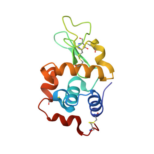
    5OLD, 5OLE

  • PubMed Abstract: 

    The tetranuclear Pt complex (PtL) 4 (where L 2- is the anion derived from para-isopropyl thiosemicarbazone) was first described in A.G. Quiroga et al., J. Med. Chem. 41, 1998, 1399-1408. (PtL) 4 manifests antiproliferative properties toward various cancer cell lines being a promising anticancer drug candidate. Yet, details of its reactivity with biomolecules have not been elucidated. To this end, we investigated the reactions of (PtL) 4 with a few model proteins, i.e. bovine pancreatic ribonuclease (RNase A), cytochrome c (Cyt c) and hen egg white lysozyme (Lysozyme), through electrospray ionization mass spectrometry and other biophysical methods. A rich reactivity of (PtL) 4 with the above-mentioned model proteins is observed, leading to the formation of numerous metallodrug-protein adducts. The tetranuclear complex breaks down and various fragments bind proteins up to high metal/protein ratios; this typically results into very complicated mass spectral patterns. However, some of the main mass peaks could be assigned in the case of the Lysozyme adduct. In addition, crystallographic data were obtained for the (PtL) 4 /Lysozyme and (PtL) 4 /RNase A adducts pointing at His side chains as the primary binding sites for monometallic Pt fragments. Notably, a few selected features of the interactions observed in the (PtL) 4 /protein adducts were reproduced by reacting (PtL) 4 with a small molecule, i.e. N-methylimidazole. In conclusion, the present study confirms the prodrug nature of the tetraplatinum complex, clarifies one possible pathway for its activation through cluster disassembly and allows initial identification of adducts formed with a representative protein.


  • Organizational Affiliation
    • Department of Chemistry and Industrial Chemistry (DCCI), University of Pisa, Via Moruzzi, 13, 56124 Pisa, Italy; Laboratory of Metals in Medicine (MetMed), Department of Chemistry "U. Schiff", University of Florence, Via della Lastruccia 3, 50019 Sesto Fiorentino, Italy.

Macromolecule Content 

  • Total Structure Weight: 15.45 kDa 
  • Atom Count: 1,166 
  • Modeled Residue Count: 129 
  • Deposited Residue Count: 129 
  • Unique protein chains: 1

Macromolecules

Find similar proteins by:|  3D Structure
Entity ID: 1
MoleculeChains  Sequence LengthOrganismDetailsImage
Lysozyme C129Gallus gallusMutation(s): 0 
EC: 3.2.1.17
UniProt
Find proteins for P00698 (Gallus gallus)
Explore P00698 
Go to UniProtKB:  P00698
Entity Groups
Sequence Clusters30% Identity50% Identity70% Identity90% Identity95% Identity100% Identity
UniProt GroupP00698
Sequence Annotations
Expand
Reference Sequence

Small Molecules

Ligands 4 Unique
IDChains Name / Formula / InChI Key2D Diagram3D Interactions
PT
(Subject of Investigation/LOI)

Query on PT


Download:Ideal Coordinates CCD File
L [auth A],
M [auth A]
PLATINUM (II) ION
Pt
HRGDZIGMBDGFTC-UHFFFAOYSA-N
DMS

Query on DMS


Download:Ideal Coordinates CCD File
I [auth A],
J [auth A],
K [auth A]
DIMETHYL SULFOXIDE
C2 H6 O S
IAZDPXIOMUYVGZ-UHFFFAOYSA-N
EDO

Query on EDO


Download:Ideal Coordinates CCD File
B [auth A],
N [auth A]
1,2-ETHANEDIOL
C2 H6 O2
LYCAIKOWRPUZTN-UHFFFAOYSA-N
NO3

Query on NO3


Download:Ideal Coordinates CCD File
C [auth A]
D [auth A]
E [auth A]
F [auth A]
G [auth A]
C [auth A],
D [auth A],
E [auth A],
F [auth A],
G [auth A],
H [auth A]
NITRATE ION
N O3
NHNBFGGVMKEFGY-UHFFFAOYSA-N

Experimental Data & Validation

Experimental Data

  • Method: X-RAY DIFFRACTION
  • Resolution: 1.78 Å
  • R-Value Free:  0.238 (Depositor), 0.253 (DCC) 
  • R-Value Work:  0.181 (Depositor), 0.189 (DCC) 
  • R-Value Observed: 0.184 (Depositor) 
Space Group: P 43 21 2
Unit Cell:
Length ( Å )Angle ( ˚ )
a = 77.387α = 90
b = 77.387β = 90
c = 37.626γ = 90
Software Package:
Software NamePurpose
REFMACrefinement
HKL-2000data reduction
HKL-2000data scaling
PHASERphasing

Structure Validation

View Full Validation Report


Entry History 

Deposition Data

Revision History  (Full details and data files)

  • Version 1.0: 2018-08-29
    Type: Initial release
  • Version 1.1: 2019-03-13
    Changes: Data collection, Database references
  • Version 1.2: 2024-01-17
    Changes: Data collection, Database references, Derived calculations, Refinement description
  • Version 1.3: 2024-10-16
    Changes: Structure summary