5OJJ | pdb_00005ojj

Crystal structure of the Zn-bound ubiquitin-conjugating enzyme Ube2T


Experimental Data Snapshot

  • Method: X-RAY DIFFRACTION
  • Resolution: 1.85 Å
  • R-Value Free: 
    0.199 (Depositor), 0.207 (DCC) 
  • R-Value Work: 
    0.178 (Depositor), 0.187 (DCC) 
  • R-Value Observed: 
    0.179 (Depositor) 

Starting Model: experimental
View more details

wwPDB Validation 3D Report Full Report

Validation slider image for 5OJJ

This is version 1.2 of the entry. See complete history

Literature

Mind the Metal: A Fragment Library-Derived Zinc Impurity Binds the E2 Ubiquitin-Conjugating Enzyme Ube2T and Induces Structural Rearrangements.

Morreale, F.E.Testa, A.Chaugule, V.K.Bortoluzzi, A.Ciulli, A.Walden, H.

(2017) J Med Chem 60: 8183-8191

  • DOI: https://doi.org/10.1021/acs.jmedchem.7b01071
  • Primary Citation Related Structures: 
    5OJJ

  • PubMed Abstract: 

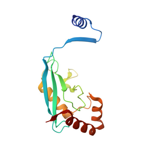
    Efforts to develop inhibitors, activators, and effectors of biological reactions using small molecule libraries are often hampered by interference compounds, artifacts, and false positives that permeate the pool of initial hits. Here, we report the discovery of a promising initial hit compound targeting the Fanconi anemia ubiquitin-conjugating enzyme Ube2T and describe its biophysical and biochemical characterization. Analysis of the co-crystal structure led to the identification of a contaminating zinc ion as solely responsible for the observed effects. Zinc binding to the active site cysteine induces a domain swap in Ube2T that leads to cyclic trimerization organized in an open-ended linear assembly. Our study serves as a cautionary tale for screening small molecule libraries and provides insights into the structural plasticity of ubiquitin-conjugating enzymes.


  • Organizational Affiliation
    • MRC Protein Phosphorylation and Ubiquitylation Unit, ‡Division of Biological Chemistry and Drug Discovery, School of Life Sciences, University of Dundee , Dundee DD1 5EH, United Kingdom.

Macromolecule Content 

  • Total Structure Weight: 107.24 kDa 
  • Atom Count: 8,268 
  • Modeled Residue Count: 904 
  • Deposited Residue Count: 924 
  • Unique protein chains: 1

Macromolecules

Find similar proteins by:|  3D Structure
Entity ID: 1
MoleculeChains  Sequence LengthOrganismDetailsImage
Ubiquitin-conjugating enzyme E2 T
A, B, C, D, E
A, B, C, D, E, F
154Homo sapiensMutation(s): 0 
Gene Names: UBE2THSPC150PIG50
EC: 2.3.2.23
UniProt & NIH Common Fund Data Resources
Find proteins for Q9NPD8 (Homo sapiens)
Explore Q9NPD8 
Go to UniProtKB:  Q9NPD8
PHAROS:  Q9NPD8
GTEx:  ENSG00000077152 
Entity Groups
Sequence Clusters30% Identity50% Identity70% Identity90% Identity95% Identity100% Identity
UniProt GroupQ9NPD8
Sequence Annotations
Expand
Reference Sequence

Small Molecules

Ligands 3 Unique
IDChains Name / Formula / InChI Key2D Diagram3D Interactions
TRS

Query on TRS


Download:Ideal Coordinates CCD File
I [auth A],
O [auth C]
2-AMINO-2-HYDROXYMETHYL-PROPANE-1,3-DIOL
C4 H12 N O3
LENZDBCJOHFCAS-UHFFFAOYSA-O
ZN

Query on ZN


Download:Ideal Coordinates CCD File
H [auth A]
J [auth A]
L [auth B]
N [auth C]
Q [auth D]
H [auth A],
J [auth A],
L [auth B],
N [auth C],
Q [auth D],
R [auth D],
T [auth E],
V [auth F]
ZINC ION
Zn
PTFCDOFLOPIGGS-UHFFFAOYSA-N
ACT

Query on ACT


Download:Ideal Coordinates CCD File
G [auth A]
K [auth B]
M [auth C]
P [auth D]
S [auth E]
G [auth A],
K [auth B],
M [auth C],
P [auth D],
S [auth E],
U [auth F]
ACETATE ION
C2 H3 O2
QTBSBXVTEAMEQO-UHFFFAOYSA-M

Experimental Data & Validation

Experimental Data

  • Method: X-RAY DIFFRACTION
  • Resolution: 1.85 Å
  • R-Value Free:  0.199 (Depositor), 0.207 (DCC) 
  • R-Value Work:  0.178 (Depositor), 0.187 (DCC) 
  • R-Value Observed: 0.179 (Depositor) 
Space Group: P 1 21 1
Unit Cell:
Length ( Å )Angle ( ˚ )
a = 56.02α = 90
b = 96.383β = 93.28
c = 89.996γ = 90
Software Package:
Software NamePurpose
REFMACrefinement
XDSdata reduction
Aimlessdata scaling
MOLREPphasing

Structure Validation

View Full Validation Report


Entry History 

Deposition Data

Revision History  (Full details and data files)

  • Version 1.0: 2017-10-04
    Type: Initial release
  • Version 1.1: 2017-10-25
    Changes: Database references
  • Version 1.2: 2024-01-17
    Changes: Data collection, Database references, Derived calculations, Refinement description