5OHX | pdb_00005ohx

Structure of active cystathionine B-synthase from Apis mellifera


Experimental Data Snapshot

  • Method: X-RAY DIFFRACTION
  • Resolution: 3.20 Å
  • R-Value Free: 
    0.231 (Depositor), 0.231 (DCC) 
  • R-Value Work: 
    0.197 (Depositor), 0.197 (DCC) 
  • R-Value Observed: 
    0.199 (Depositor) 

Starting Model: experimental
View more details

wwPDB Validation 3D Report Full Report

Validation slider image for 5OHX

Ligand Structure Quality Assessment 


This is version 1.3 of the entry. See complete history

Literature

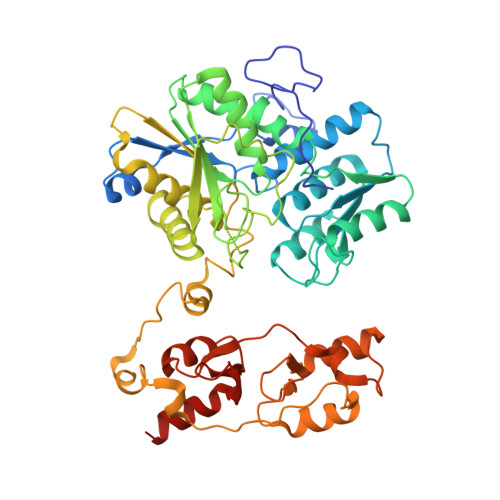
Crystal structure of cystathionine beta-synthase from honeybee Apis mellifera.

Gimenez-Mascarell, P.Majtan, T.Oyenarte, I.Ereno-Orbea, J.Majtan, J.Klaudiny, J.Kraus, J.P.Martinez-Cruz, L.A.

(2018) J Struct Biol 202: 82-93

  • DOI: https://doi.org/10.1016/j.jsb.2017.12.008
  • Primary Citation Related Structures: 
    5OHX

  • PubMed Abstract: 

    Cystathionine β-synthase (CBS), the key enzyme in the transsulfuration pathway, links methionine metabolism to the biosynthesis of cellular redox controlling molecules. CBS catalyzes the pyridoxal-5'-phosphate-dependent condensation of serine and homocysteine to form cystathionine, which is subsequently converted into cysteine. Besides maintaining cellular sulfur amino acid homeostasis, CBS also catalyzes multiple hydrogen sulfide-generating reactions using cysteine and homocysteine as substrates. In mammals, CBS is activated by S-adenosylmethionine (AdoMet), where it can adopt two different conformations (basal and activated), but exists as a unique highly active species in fruit fly Drosophila melanogaster. Here we present the crystal structure of CBS from honeybey Apis mellifera, which shows a constitutively active dimeric species and let explain why the enzyme is not allosterically regulated by AdoMet. In addition, comparison of available CBS structures unveils a substrate-induced closure of the catalytic cavity, which in humans is affected by the AdoMet-dependent regulation and likely impaired by the homocystinuria causing mutation T191M.


  • Organizational Affiliation
    • Structural Biology Unit, Center for Cooperative Research in Biosciences (CIC Biogune), Technology Park of Bizkaia, 48160 Derio, Bizkaia, Spain.

Macromolecule Content 

  • Total Structure Weight: 114.3 kDa 
  • Atom Count: 7,706 
  • Modeled Residue Count: 976 
  • Deposited Residue Count: 1,008 
  • Unique protein chains: 1

Macromolecules

Find similar proteins by:|  3D Structure
Entity ID: 1
MoleculeChains  Sequence LengthOrganismDetailsImage
Cystathionine beta-synthase
A, B
504Apis melliferaMutation(s): 0 
Gene Names: AmCBS
EC: 4.2.1.22
UniProt
Find proteins for Q2V0C9 (Apis mellifera)
Explore Q2V0C9 
Go to UniProtKB:  Q2V0C9
Entity Groups
Sequence Clusters30% Identity50% Identity70% Identity90% Identity95% Identity100% Identity
UniProt GroupQ2V0C9
Sequence Annotations
Expand
Reference Sequence

Experimental Data & Validation

Experimental Data

  • Method: X-RAY DIFFRACTION
  • Resolution: 3.20 Å
  • R-Value Free:  0.231 (Depositor), 0.231 (DCC) 
  • R-Value Work:  0.197 (Depositor), 0.197 (DCC) 
  • R-Value Observed: 0.199 (Depositor) 
Space Group: P 21 21 21
Unit Cell:
Length ( Å )Angle ( ˚ )
a = 86.143α = 90
b = 96.099β = 90
c = 180.684γ = 90
Software Package:
Software NamePurpose
PHENIXrefinement
HKL-2000data reduction
Aimlessdata scaling
PHENIXphasing
Cootmodel building

Structure Validation

View Full Validation Report


Ligand Structure Quality Assessment 


Entry History 

Deposition Data

Revision History  (Full details and data files)

  • Version 1.0: 2018-01-03
    Type: Initial release
  • Version 1.1: 2018-03-14
    Changes: Database references
  • Version 1.2: 2019-10-09
    Changes: Data collection
  • Version 1.3: 2024-01-17
    Changes: Data collection, Database references, Derived calculations, Refinement description