5OHE | pdb_00005ohe

Globin sensor domain of AfGcHK (FeIII form) in complex with cyanide


Experimental Data Snapshot

  • Method: X-RAY DIFFRACTION
  • Resolution: 1.85 Å
  • R-Value Free: 
    0.241 (Depositor), 0.205 (DCC) 
  • R-Value Work: 
    0.192 (Depositor), 0.202 (DCC) 
  • R-Value Observed: 
    0.194 (Depositor) 

Starting Model: experimental
View more details

wwPDB Validation 3D Report Full Report

Validation slider image for 5OHE

Ligand Structure Quality Assessment 


This is version 1.4 of the entry. See complete history

Literature

Coordination and redox state-dependent structural changes of the heme-based oxygen sensor AfGcHK associated with intraprotein signal transduction.

Stranava, M.Man, P.Skalova, T.Kolenko, P.Blaha, J.Fojtikova, V.Martinek, V.Dohnalek, J.Lengalova, A.Rosulek, M.Shimizu, T.Martinkova, M.

(2017) J Biological Chem 292: 20921-20935

  • DOI: https://doi.org/10.1074/jbc.M117.817023
  • Primary Citation Related Structures: 
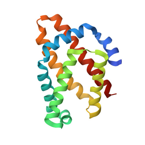
    5OHE, 5OHF

  • PubMed Abstract: 

    The heme-based oxygen sensor histidine kinase Af GcHK is part of a two-component signal transduction system in bacteria. O 2 binding to the Fe(II) heme complex of its N-terminal globin domain strongly stimulates autophosphorylation at His 183 in its C-terminal kinase domain. The 6-coordinate heme Fe(III)-OH - and -CN - complexes of Af GcHK are also active, but the 5-coordinate heme Fe(II) complex and the heme-free apo-form are inactive. Here, we determined the crystal structures of the isolated dimeric globin domains of the active Fe(III)-CN - and inactive 5-coordinate Fe(II) forms, revealing striking structural differences on the heme-proximal side of the globin domain. Using hydrogen/deuterium exchange coupled with mass spectrometry to characterize the conformations of the active and inactive forms of full-length Af GcHK in solution, we investigated the intramolecular signal transduction mechanisms. Major differences between the active and inactive forms were observed on the heme-proximal side (helix H5), at the dimerization interface (helices H6 and H7 and loop L7) of the globin domain and in the ATP-binding site (helices H9 and H11) of the kinase domain. Moreover, separation of the sensor and kinase domains, which deactivates catalysis, increased the solvent exposure of the globin domain-dimerization interface (helix H6) as well as the flexibility and solvent exposure of helix H11. Together, these results suggest that structural changes at the heme-proximal side, the globin domain-dimerization interface, and the ATP-binding site are important in the signal transduction mechanism of Af GcHK. We conclude that Af GcHK functions as an ensemble of molecules sampling at least two conformational states.


  • Organizational Affiliation
    • From the Department of Biochemistry, Faculty of Science, Charles University, Hlavova (Albertov) 2030/8, Prague 2, 128 43 Czech Republic.

Macromolecule Content 

  • Total Structure Weight: 152.22 kDa 
  • Atom Count: 11,460 
  • Modeled Residue Count: 1,218 
  • Deposited Residue Count: 1,288 
  • Unique protein chains: 1

Macromolecules

Find similar proteins by:|  3D Structure
Entity ID: 1
MoleculeChains  Sequence LengthOrganismDetailsImage
Globin-coupled histidine kinase
A, B, C, D, E
A, B, C, D, E, F, G, H
161Anaeromyxobacter sp. Fw109-5Mutation(s): 0 
Gene Names: gchKAnae109_2438
EC: 2.7.13.3
UniProt
Find proteins for A7HD43 (Anaeromyxobacter sp. (strain Fw109-5))
Explore A7HD43 
Go to UniProtKB:  A7HD43
Entity Groups
Sequence Clusters30% Identity50% Identity70% Identity90% Identity95% Identity100% Identity
UniProt GroupA7HD43
Sequence Annotations
Expand
Reference Sequence

Small Molecules

Ligands 4 Unique
IDChains Name / Formula / InChI Key2D Diagram3D Interactions
HEM

Query on HEM


Download:Ideal Coordinates CCD File
I [auth A]
L [auth B]
N [auth C]
Q [auth D]
R [auth E]
I [auth A],
L [auth B],
N [auth C],
Q [auth D],
R [auth E],
V [auth F],
W [auth G],
Z [auth H]
PROTOPORPHYRIN IX CONTAINING FE
C34 H32 Fe N4 O4
KABFMIBPWCXCRK-RGGAHWMASA-L
CL

Query on CL


Download:Ideal Coordinates CCD File
K [auth B],
P [auth D],
T [auth E],
Y [auth G]
CHLORIDE ION
Cl
VEXZGXHMUGYJMC-UHFFFAOYSA-M
CYN

Query on CYN


Download:Ideal Coordinates CCD File
AA [auth H]
J [auth A]
M [auth B]
O [auth C]
S [auth E]
AA [auth H],
J [auth A],
M [auth B],
O [auth C],
S [auth E],
X [auth G]
CYANIDE ION
C N
XFXPMWWXUTWYJX-UHFFFAOYSA-N
NA

Query on NA


Download:Ideal Coordinates CCD File
U [auth E]SODIUM ION
Na
FKNQFGJONOIPTF-UHFFFAOYSA-N

Experimental Data & Validation

Experimental Data

  • Method: X-RAY DIFFRACTION
  • Resolution: 1.85 Å
  • R-Value Free:  0.241 (Depositor), 0.205 (DCC) 
  • R-Value Work:  0.192 (Depositor), 0.202 (DCC) 
  • R-Value Observed: 0.194 (Depositor) 
Space Group: P 41 21 2
Unit Cell:
Length ( Å )Angle ( ˚ )
a = 78.069α = 90
b = 78.069β = 90
c = 441.819γ = 90
Software Package:
Software NamePurpose
REFMACrefinement
XDSdata reduction
Aimlessdata scaling
MoRDaphasing

Structure Validation

View Full Validation Report


Ligand Structure Quality Assessment 


Entry History 

& Funding Information

Deposition Data


Funding OrganizationLocationGrant Number
Czech Science FoundationCzech Republic15-19883S
Ministry of Education, Youth and Sports of the Czech RepublicCzech RepublicCZ.1.05/1.1.00/02.109
Grant Agency of the Czech Technical UniversityCzech RepublicSGS16/246/OHK4/3T/14
Ministry of Education, Youth and Sports of the Czech RepublicCzech RepublicLM2015043
Ministry of Education, Youth and Sports of the Czech RepublicCzech RepublicCZ.02.1.01/0.0/0.0/16_013/0001776

Revision History  (Full details and data files)

  • Version 1.0: 2017-11-08
    Type: Initial release
  • Version 1.1: 2017-11-15
    Changes: Database references
  • Version 1.2: 2018-01-03
    Changes: Database references
  • Version 1.3: 2018-01-10
    Changes: Source and taxonomy
  • Version 1.4: 2024-01-17
    Changes: Data collection, Database references, Derived calculations, Refinement description