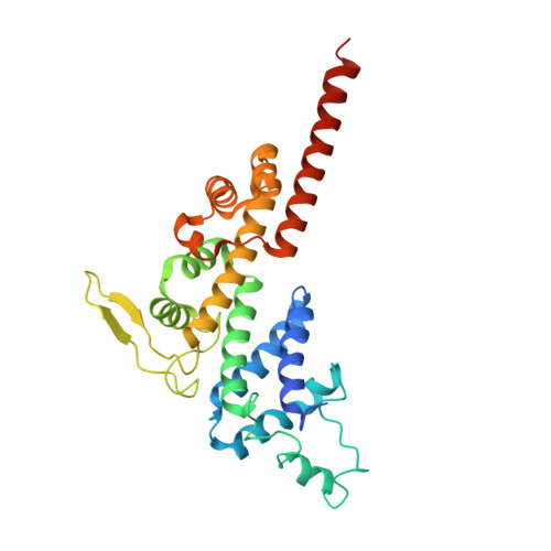
5OH5 | pdb_00005oh5

Legionella pneumophila RidL N-terminal retromer binding domain


Experimental Data Snapshot

  • Method: X-RAY DIFFRACTION
  • Resolution: 1.90 Å
  • R-Value Free: 
    0.214 (Depositor), 0.217 (DCC) 
  • R-Value Work: 
    0.204 (Depositor), 0.210 (DCC) 
  • R-Value Observed: 
    0.204 (Depositor) 

wwPDB Validation 3D Report Full Report

Validation slider image for 5OH5

This is version 1.2 of the entry. See complete history. 

Literature

Structural insights into Legionella RidL-Vps29 retromer subunit interaction reveal displacement of the regulator TBC1D5.

Barlocher, K., Hutter, C.A.J., Swart, A.L., Steiner, B., Welin, A., Hohl, M., Letourneur, F., Seeger, M.A., Hilbi, H.

(2017) Nat Commun 8: 1543-1543

  • DOI: https://doi.org/10.1038/s41467-017-01512-5
  • Primary Citation Related Structures: 
    5OH5, 5OH6

  • PubMed Abstract: 

    Legionella pneumophila can cause Legionnaires' disease and replicates intracellularly in a distinct Legionella-containing vacuole (LCV). LCV formation is a complex process that involves a plethora of type IV-secreted effector proteins. The effector RidL binds the Vps29 retromer subunit, blocks retrograde vesicle trafficking, and promotes intracellular bacterial replication. Here, we reveal that the 29-kDa N-terminal domain of RidL (RidL 2-281 ) adopts a "foot-like" fold comprising a protruding β-hairpin at its "heel". The deletion of the β-hairpin, the exchange to Glu of Ile 170 in the β-hairpin, or Leu 152 in Vps29 abolishes the interaction in eukaryotic cells and in vitro. RidL 2-281 or RidL displace the Rab7 GTPase-activating protein (GAP) TBC1D5 from the retromer and LCVs, respectively, and TBC1D5 promotes the intracellular growth of L. pneumophila. Thus, the hydrophobic β-hairpin of RidL is critical for binding of the L. pneumophila effector to the Vps29 retromer subunit and displacement of the regulator TBC1D5.


  • Organizational Affiliation: 
    • Institute of Medical Microbiology, University of Zürich, Gloriastrasse 30, 8006, Zürich, Switzerland.

Macromolecule Content 

  • Total Structure Weight: 32.09 kDa 
  • Atom Count: 2,333 
  • Modeled Residue Count: 268 
  • Deposited Residue Count: 283 
  • Unique protein chains: 1

Macromolecules

Find similar proteins by:|  3D Structure
Entity ID: 1
MoleculeChains  Sequence LengthOrganismDetailsImage
RidL283Legionella pneumophila subsp. pneumophila ATCC 43290Mutation(s): 0 
Gene Names: lp12_2303
UniProt
Find proteins for A0ABF7PFW6 (Legionella pneumophila subsp. pneumophila ATCC 43290)
Explore A0ABF7PFW6 
Go to UniProtKB:  A0ABF7PFW6
Entity Groups
Sequence Clusters30% Identity50% Identity70% Identity90% Identity95% Identity100% Identity
UniProt GroupA0ABF7PFW6
Sequence Annotations
Expand
Reference Sequence

Experimental Data & Validation

Experimental Data

  • Method: X-RAY DIFFRACTION
  • Resolution: 1.90 Å
  • R-Value Free:  0.214 (Depositor), 0.217 (DCC) 
  • R-Value Work:  0.204 (Depositor), 0.210 (DCC) 
  • R-Value Observed: 0.204 (Depositor) 
Space Group: I 4 2 2
Unit Cell:
Length ( Å )Angle ( ˚ )
a = 110.14α = 90
b = 110.14β = 90
c = 98.51γ = 90
Software Package:
Software NamePurpose
XSCALEdata scaling
PHENIXrefinement
PDB_EXTRACTdata extraction
XDSdata reduction
PHASERphasing

Structure Validation

View Full Validation Report


Entry History 

Deposition Data

Revision History  (Full details and data files)

  • Version 1.0: 2017-11-22
    Type: Initial release
  • Version 1.1: 2017-11-29
    Changes: Database references
  • Version 1.2: 2024-05-08
    Changes: Data collection, Database references