5OG2 | pdb_00005og2

Cu nitrite reductase serial data at varying temperatures RT 0.09MGy


Experimental Data Snapshot

  • Method: X-RAY DIFFRACTION
  • Resolution: 1.64 Å
  • R-Value Free: 
    0.190 (Depositor), 0.190 (DCC) 
  • R-Value Work: 
    0.164 (Depositor) 
  • R-Value Observed: 
    0.165 (Depositor) 

Starting Model: experimental
View more details

wwPDB Validation 3D Report Full Report

Validation slider image for 5OG2

This is version 1.1 of the entry. See complete history

Literature

Enzyme catalysis captured using multiple structures from one crystal at varying temperatures.

Horrell, S.Kekilli, D.Sen, K.Owen, R.L.Dworkowski, F.S.N.Antonyuk, S.V.Keal, T.W.Yong, C.W.Eady, R.R.Hasnain, S.S.Strange, R.W.Hough, M.A.

(2018) IUCrJ 5: 283-292

  • DOI: https://doi.org/10.1107/S205225251800386X
  • Primary Citation Related Structures: 
    5OF5, 5OF6, 5OF7, 5OF8, 5OFC, 5OFD, 5OFE, 5OFF, 5OFG, 5OFH, 5OG2, 5OG3, 5OG4, 5OG5, 5OG6, 5OGF, 5OGG

  • PubMed Abstract: 

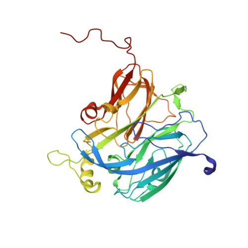
    High-resolution crystal structures of enzymes in relevant redox states have transformed our understanding of enzyme catalysis. Recent developments have demonstrated that X-rays can be used, via the generation of solvated electrons, to drive reactions in crystals at cryogenic temperatures (100 K) to generate 'structural movies' of enzyme reactions. However, a serious limitation at these temperatures is that protein conformational motion can be significantly supressed. Here, the recently developed MSOX (multiple serial structures from one crystal) approach has been applied to nitrite-bound copper nitrite reductase at room temperature and at 190 K, close to the glass transition. During both series of multiple structures, nitrite was initially observed in a 'top-hat' geometry, which was rapidly transformed to a 'side-on' configuration before conversion to side-on NO, followed by dissociation of NO and substitution by water to reform the resting state. Density functional theory calculations indicate that the top-hat orientation corresponds to the oxidized type 2 copper site, while the side-on orientation is consistent with the reduced state. It is demonstrated that substrate-to-product conversion within the crystal occurs at a lower radiation dose at 190 K, allowing more of the enzyme catalytic cycle to be captured at high resolution than in the previous 100 K experiment. At room temperature the reaction was very rapid, but it remained possible to generate and characterize several structural states. These experiments open up the possibility of obtaining MSOX structural movies at multiple temperatures (MSOX-VT), providing an unparallelled level of structural information during catalysis for redox enzymes.


  • Organizational Affiliation
    • School of Biological Sciences, University of Essex, Wivenhoe Park, Colchester CO4 3SQ, England.

Macromolecule Content 

  • Total Structure Weight: 37.08 kDa 
  • Atom Count: 2,800 
  • Modeled Residue Count: 332 
  • Deposited Residue Count: 334 
  • Unique protein chains: 1

Macromolecules

Find similar proteins by:|  3D Structure
Entity ID: 1
MoleculeChains  Sequence LengthOrganismDetailsImage
Copper-containing nitrite reductase334Achromobacter cycloclastesMutation(s): 0 
Gene Names: nirK
EC: 1.7.2.1
UniProt
Find proteins for P25006 (Achromobacter cycloclastes)
Explore P25006 
Go to UniProtKB:  P25006
Entity Groups
Sequence Clusters30% Identity50% Identity70% Identity90% Identity95% Identity100% Identity
UniProt GroupP25006
Sequence Annotations
Expand
Reference Sequence

Small Molecules

Ligands 3 Unique
IDChains Name / Formula / InChI Key2D Diagram3D Interactions
CU
(Subject of Investigation/LOI)

Query on CU


Download:Ideal Coordinates CCD File
B [auth A],
C [auth A]
COPPER (II) ION
Cu
JPVYNHNXODAKFH-UHFFFAOYSA-N
ACT

Query on ACT


Download:Ideal Coordinates CCD File
E [auth A]ACETATE ION
C2 H3 O2
QTBSBXVTEAMEQO-UHFFFAOYSA-M
NO2
(Subject of Investigation/LOI)

Query on NO2


Download:Ideal Coordinates CCD File
D [auth A]
F [auth A]
G [auth A]
H [auth A]
I [auth A]
D [auth A],
F [auth A],
G [auth A],
H [auth A],
I [auth A],
J [auth A]
NITRITE ION
N O2
IOVCWXUNBOPUCH-UHFFFAOYSA-M

Experimental Data & Validation

Experimental Data

  • Method: X-RAY DIFFRACTION
  • Resolution: 1.64 Å
  • R-Value Free:  0.190 (Depositor), 0.190 (DCC) 
  • R-Value Work:  0.164 (Depositor) 
  • R-Value Observed: 0.165 (Depositor) 
Space Group: P 21 3
Unit Cell:
Length ( Å )Angle ( ˚ )
a = 96.241α = 90
b = 96.241β = 90
c = 96.241γ = 90
Software Package:
Software NamePurpose
REFMACrefinement
DIALSdata collection
XDSdata reduction
Aimlessdata scaling
REFMACphasing

Structure Validation

View Full Validation Report


Entry History 

& Funding Information

Deposition Data


Funding OrganizationLocationGrant Number
Biotechnology and Biological Sciences Research CouncilUnited KingdomBB/M022714/1
Biotechnology and Biological Sciences Research CouncilUnited KingdomBB/M020924/1
Leverhulme TrustUnited KingdomRPG-2014-355

Revision History  (Full details and data files)

  • Version 1.0: 2018-05-23
    Type: Initial release
  • Version 1.1: 2024-01-17
    Changes: Data collection, Database references, Derived calculations, Refinement description