5OF0 | pdb_00005of0

X-ray structure of human glutamate carboxypeptidase II (GCPII), the E424M inactive mutant, in complex with a inhibitor CFBzOG


Experimental Data Snapshot

  • Method: X-RAY DIFFRACTION
  • Resolution: 1.48 Å
  • R-Value Free: 
    0.172 (Depositor), 0.179 (DCC) 
  • R-Value Work: 
    0.157 (Depositor), 0.164 (DCC) 
  • R-Value Observed: 
    0.157 (Depositor) 

Starting Model: experimental
View more details

wwPDB Validation 3D Report Full Report

Validation slider image for 5OF0

Ligand Structure Quality Assessment 


This is version 2.2 of the entry. See complete history

Literature

2-Aminoadipic Acid-C(O)-Glutamate Based Prostate-Specific Membrane Antigen Ligands for Potential Use as Theranostics.

Nakajima, R.Novakova, Z.Tueckmantel, W.Motlova, L.Barinka, C.Kozikowski, A.P.

(2018) ACS Med Chem Lett 9: 1099-1104

  • DOI: https://doi.org/10.1021/acsmedchemlett.8b00318
  • Primary Citation Related Structures: 
    5OF0, 6H7Y, 6H7Z, 6HKJ, 6HKZ

  • PubMed Abstract: 

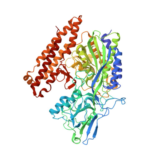
    The design and synthesis of prostate specific membrane antigen (PSMA) ligands derived from 2-aminoadipic acid, a building block that has not previously been used to construct PSMA ligands, are reported. The effects of both the linker length and of an N-substituent of our PSMA ligands were probed, and X-ray structures of five of these ligands bound to PSMA were obtained. Among the ligands disclosed herein, 13b showed the highest inhibitory activity for PSMA. As ligand 13b can readily be radiolabeled since its fluorine atom is adjacent to the nitrogen atom of its pyridine ring, the use of this and related compounds as theranostics can be pursued.


  • Organizational Affiliation
    • Department of Medicinal Chemistry and Pharmacognosy, University of Illinois at Chicago, Chicago, Illinois 60612, United States.

Macromolecule Content 

  • Total Structure Weight: 83.64 kDa 
  • Atom Count: 6,629 
  • Modeled Residue Count: 692 
  • Deposited Residue Count: 707 
  • Unique protein chains: 1

Macromolecules

Find similar proteins by:|  3D Structure
Entity ID: 1
MoleculeChains  Sequence LengthOrganismDetailsImage
Glutamate carboxypeptidase 2707Homo sapiensMutation(s): 1 
Gene Names: FOLH1FOLHNAALAD1PSMPSMAGIG27
EC: 3.4.17.21
UniProt & NIH Common Fund Data Resources
Find proteins for Q04609 (Homo sapiens)
Explore Q04609 
Go to UniProtKB:  Q04609
PHAROS:  Q04609
GTEx:  ENSG00000086205 
Entity Groups
Sequence Clusters30% Identity50% Identity70% Identity90% Identity95% Identity100% Identity
UniProt GroupQ04609
Glycosylation
Glycosylation Sites: 7Go to GlyGen: Q04609-1
Sequence Annotations
Expand
Reference Sequence

Oligosaccharides

Help  
Entity ID: 2
MoleculeChains Length2D Diagram GlycosylationD Interactions
2-acetamido-2-deoxy-beta-D-glucopyranose-(1-4)-2-acetamido-2-deoxy-beta-D-glucopyranose
B, C, D
2N-Glycosylation
Glycosylation Resources
GlyTouCan: G42666HT
GlyCosmos: G42666HT
GlyGen: G42666HT
Entity ID: 3
MoleculeChains Length2D Diagram GlycosylationD Interactions
alpha-D-mannopyranose-(1-3)-beta-D-mannopyranose-(1-4)-2-acetamido-2-deoxy-beta-D-glucopyranose-(1-4)-2-acetamido-2-deoxy-beta-D-glucopyranose
E
4N-Glycosylation
Glycosylation Resources
GlyTouCan: G81315DD
GlyCosmos: G81315DD
GlyGen: G81315DD
Entity ID: 4
MoleculeChains Length2D Diagram GlycosylationD Interactions
alpha-D-mannopyranose-(1-3)-[alpha-D-mannopyranose-(1-6)]beta-D-mannopyranose-(1-4)-2-acetamido-2-deoxy-beta-D-glucopyranose-(1-4)-2-acetamido-2-deoxy-beta-D-glucopyranose
F
5N-Glycosylation
Glycosylation Resources
GlyTouCan: G22768VO
GlyCosmos: G22768VO
GlyGen: G22768VO

Small Molecules

Ligands 5 Unique
IDChains Name / Formula / InChI Key2D Diagram3D Interactions
9TB

Query on 9TB


Download:Ideal Coordinates CCD File
M [auth A](2~{S})-2-[[(2~{S})-6-[(4-fluorophenyl)methylamino]-1-oxidanyl-1,6-bis(oxidanylidene)hexan-2-yl]carbamoylamino]pentanedioic acid
C19 H24 F N3 O8
ZCBCSGACSCSXNO-KBPBESRZSA-N
NAG

Query on NAG


Download:Ideal Coordinates CCD File
G [auth A],
H [auth A]
2-acetamido-2-deoxy-beta-D-glucopyranose
C8 H15 N O6
OVRNDRQMDRJTHS-FMDGEEDCSA-N
ZN

Query on ZN


Download:Ideal Coordinates CCD File
I [auth A],
J [auth A]
ZINC ION
Zn
PTFCDOFLOPIGGS-UHFFFAOYSA-N
CA

Query on CA


Download:Ideal Coordinates CCD File
K [auth A]CALCIUM ION
Ca
BHPQYMZQTOCNFJ-UHFFFAOYSA-N
CL

Query on CL


Download:Ideal Coordinates CCD File
L [auth A]CHLORIDE ION
Cl
VEXZGXHMUGYJMC-UHFFFAOYSA-M
Binding Affinity Annotations 
IDSourceBinding Affinity
9TB BindingDB:  5OF0 IC50: min: 2.3, max: 1500 (nM) from 2 assay(s)

Experimental Data & Validation

Experimental Data

  • Method: X-RAY DIFFRACTION
  • Resolution: 1.48 Å
  • R-Value Free:  0.172 (Depositor), 0.179 (DCC) 
  • R-Value Work:  0.157 (Depositor), 0.164 (DCC) 
  • R-Value Observed: 0.157 (Depositor) 
Space Group: I 2 2 2
Unit Cell:
Length ( Å )Angle ( ˚ )
a = 101.467α = 90
b = 130.461β = 90
c = 159.574γ = 90
Software Package:
Software NamePurpose
REFMACrefinement
Aimlessdata scaling
REFMACphasing
Cootmodel building
XDSdata reduction

Structure Validation

View Full Validation Report


Ligand Structure Quality Assessment 


Entry History 

Deposition Data

Revision History  (Full details and data files)

  • Version 1.0: 2018-08-01
    Type: Initial release
  • Version 1.1: 2018-12-05
    Changes: Data collection, Database references
  • Version 2.0: 2020-07-29
    Type: Remediation
    Reason: Carbohydrate remediation
    Changes: Atomic model, Data collection, Derived calculations, Structure summary
  • Version 2.1: 2024-01-17
    Changes: Data collection, Database references, Derived calculations, Refinement description, Structure summary
  • Version 2.2: 2024-10-09
    Changes: Structure summary