5OE4 | pdb_00005oe4

Crystal structure of the N-terminal domain of PqsA in complex with anthraniloyl-AMP (crystal form 2)


Experimental Data Snapshot

  • Method: X-RAY DIFFRACTION
  • Resolution: 1.90 Å
  • R-Value Free: 
    0.250 (Depositor), 0.251 (DCC) 
  • R-Value Work: 
    0.209 (Depositor), 0.210 (DCC) 
  • R-Value Observed: 
    0.211 (Depositor) 

Starting Model: experimental
View more details

wwPDB Validation 3D Report Full Report

Validation slider image for 5OE4

Ligand Structure Quality Assessment 


This is version 1.2 of the entry. See complete history

Literature

Structures of the N-Terminal Domain of PqsA in Complex with Anthraniloyl- and 6-Fluoroanthraniloyl-AMP: Substrate Activation in Pseudomonas Quinolone Signal (PQS) Biosynthesis.

Witzgall, F.Ewert, W.Blankenfeldt, W.

(2017) Chembiochem 18: 2045-2055

  • DOI: https://doi.org/10.1002/cbic.201700374
  • Primary Citation Related Structures: 
    5OE3, 5OE4, 5OE5, 5OE6

  • PubMed Abstract: 

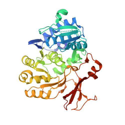
    Pseudomonas aeruginosa, a prevalent pathogen in nosocomial infections and a major burden in cystic fibrosis, uses three interconnected quorum-sensing systems to coordinate virulence processes. At variance with other Gram-negative bacteria, one of these systems relies on 2-alkyl-4(1H)-quinolones (Pseudomonas quinolone signal, PQS) and might hence be an attractive target for new anti-infective agents. Here we report crystal structures of the N-terminal domain of anthranilate-CoA ligase PqsA, the first enzyme of PQS biosynthesis, in complex with anthraniloyl-AMP and with 6-fluoroanthraniloyl-AMP (6FABA-AMP) at 1.4 and 1.7 Å resolution. We find that PqsA belongs to an unrecognized subfamily of anthranilate-CoA ligases that recognize the amino group of anthranilate through a water-mediated hydrogen bond. The complex with 6FABA-AMP explains why 6FABA, an inhibitor of PQS biosynthesis, is a good substrate of PqsA. Together, our data might pave a way to new pathoblockers in P. aeruginosa infections.


  • Organizational Affiliation
    • Structure and Function of Proteins, Helmholtz Centre for Infection Research, Inhoffenstrasse 7, 38124, Braunschweig, Germany.

Macromolecule Content 

  • Total Structure Weight: 89.72 kDa 
  • Atom Count: 6,879 
  • Modeled Residue Count: 771 
  • Deposited Residue Count: 814 
  • Unique protein chains: 1

Macromolecules

Find similar proteins by:|  3D Structure
Entity ID: 1
MoleculeChains  Sequence LengthOrganismDetailsImage
Anthranilate--CoA ligase
A, B
407Pseudomonas aeruginosa PAO1Mutation(s): 0 
Gene Names: pqsAPA0996
EC: 6.2.1.32
UniProt
Find proteins for Q9I4X3 (Pseudomonas aeruginosa (strain ATCC 15692 / DSM 22644 / CIP 104116 / JCM 14847 / LMG 12228 / 1C / PRS 101 / PAO1))
Explore Q9I4X3 
Go to UniProtKB:  Q9I4X3
Entity Groups
Sequence Clusters30% Identity50% Identity70% Identity90% Identity95% Identity100% Identity
UniProt GroupQ9I4X3
Sequence Annotations
Expand
Reference Sequence

Experimental Data & Validation

Experimental Data

  • Method: X-RAY DIFFRACTION
  • Resolution: 1.90 Å
  • R-Value Free:  0.250 (Depositor), 0.251 (DCC) 
  • R-Value Work:  0.209 (Depositor), 0.210 (DCC) 
  • R-Value Observed: 0.211 (Depositor) 
Space Group: P 1 21 1
Unit Cell:
Length ( Å )Angle ( ˚ )
a = 77.798α = 90
b = 71.966β = 116.9
c = 84.417γ = 90
Software Package:
Software NamePurpose
Aimlessdata scaling
MOLREPphasing
PHENIXrefinement
PDB_EXTRACTdata extraction
XDSdata reduction
Cootmodel building

Structure Validation

View Full Validation Report


Ligand Structure Quality Assessment 


Entry History 

Deposition Data

Revision History  (Full details and data files)

  • Version 1.0: 2017-09-06
    Type: Initial release
  • Version 1.1: 2017-10-25
    Changes: Database references
  • Version 1.2: 2024-01-17
    Changes: Data collection, Database references, Refinement description