5OD1 | pdb_00005od1

Structure of the engineered metalloesterase MID1sc10 complexed with a phosphonate transition state analogue


Experimental Data Snapshot

  • Method: X-RAY DIFFRACTION
  • Resolution: 1.34 Å
  • R-Value Free: 
    0.171 (Depositor), 0.172 (DCC) 
  • R-Value Work: 
    0.126 (Depositor), 0.127 (DCC) 
  • R-Value Observed: 
    0.129 (Depositor) 

wwPDB Validation 3D Report Full Report

Validation slider image for 5OD1

Ligand Structure Quality Assessment 


This is version 1.4 of the entry. See complete history

Literature

Evolution of a highly active and enantiospecific metalloenzyme from short peptides.

Studer, S.Hansen, D.A.Pianowski, Z.L.Mittl, P.R.E.Debon, A.Guffy, S.L.Der, B.S.Kuhlman, B.Hilvert, D.

(2018) Science 362: 1285-1288

  • DOI: https://doi.org/10.1126/science.aau3744
  • Primary Citation Related Structures: 
    5OD1, 5OD9

  • PubMed Abstract: 

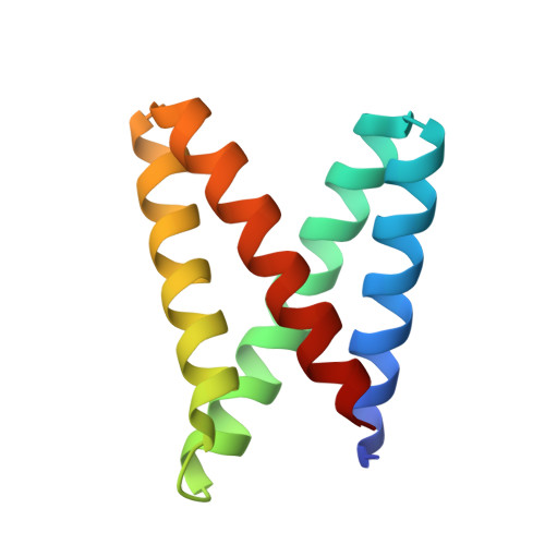
    Primordial sequence signatures in modern proteins imply ancestral origins tracing back to simple peptides. Although short peptides seldom adopt unique folds, metal ions might have templated their assembly into higher-order structures in early evolution and imparted useful chemical reactivity. Recapitulating such a biogenetic scenario, we have combined design and laboratory evolution to transform a zinc-binding peptide into a globular enzyme capable of accelerating ester cleavage with exacting enantiospecificity and high catalytic efficiency ( k cat / K M ~ 10 6 M -1 s -1 ). The simultaneous optimization of structure and function in a naïve peptide scaffold not only illustrates a plausible enzyme evolutionary pathway from the distant past to the present but also proffers exciting future opportunities for enzyme design and engineering.


  • Organizational Affiliation
    • Laboratory of Organic Chemistry, ETH Zürich, 8093 Zürich, Switzerland.

Macromolecule Content 

  • Total Structure Weight: 11.45 kDa 
  • Atom Count: 919 
  • Modeled Residue Count: 94 
  • Deposited Residue Count: 97 
  • Unique protein chains: 1

Macromolecules

Find similar proteins by:|  3D Structure
Entity ID: 1
MoleculeChains  Sequence LengthOrganismDetailsImage
MID1sc1097synthetic constructMutation(s): 0 

Small Molecules

Experimental Data & Validation

Experimental Data

  • Method: X-RAY DIFFRACTION
  • Resolution: 1.34 Å
  • R-Value Free:  0.171 (Depositor), 0.172 (DCC) 
  • R-Value Work:  0.126 (Depositor), 0.127 (DCC) 
  • R-Value Observed: 0.129 (Depositor) 
Space Group: I 2 2 2
Unit Cell:
Length ( Å )Angle ( ˚ )
a = 32.349α = 90
b = 46.877β = 90
c = 112.621γ = 90
Software Package:
Software NamePurpose
REFMACrefinement
XDSdata reduction
XSCALEdata scaling
MOLREPphasing

Structure Validation

View Full Validation Report


Ligand Structure Quality Assessment 


Entry History 

Deposition Data

Revision History  (Full details and data files)

  • Version 1.0: 2018-12-12
    Type: Initial release
  • Version 1.1: 2018-12-19
    Changes: Data collection, Structure summary
  • Version 1.2: 2019-05-29
    Changes: Data collection, Database references
  • Version 1.3: 2020-04-22
    Changes: Database references
  • Version 1.4: 2024-05-08
    Changes: Data collection, Database references