5OC7 | pdb_00005oc7

Crystal structure of the pleckstrin-homology domain of Bcr-Abl in complex with monobody Mb(Bcr-PH_4).


Experimental Data Snapshot

  • Method: X-RAY DIFFRACTION
  • Resolution: 1.65 Å
  • R-Value Free: 
    0.204 (Depositor), 0.209 (DCC) 
  • R-Value Work: 
    0.174 (Depositor) 
  • R-Value Observed: 
    0.175 (Depositor) 

Starting Model: experimental
View more details

wwPDB Validation 3D Report Full Report

Validation slider image for 5OC7

Ligand Structure Quality Assessment 


This is version 1.1 of the entry. See complete history

Literature

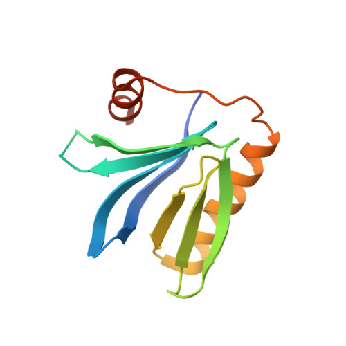
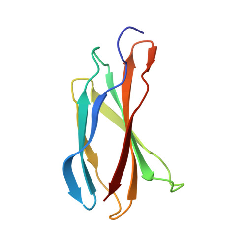
Structural and functional dissection of the DH and PH domains of oncogenic Bcr-Abl tyrosine kinase.

Reckel, S.Gehin, C.Tardivon, D.Georgeon, S.Kukenshoner, T.Lohr, F.Koide, A.Buchner, L.Panjkovich, A.Reynaud, A.Pinho, S.Gerig, B.Svergun, D.Pojer, F.Guntert, P.Dotsch, V.Koide, S.Gavin, A.C.Hantschel, O.

(2017) Nat Commun 8: 2101-2101

  • DOI: https://doi.org/10.1038/s41467-017-02313-6
  • Primary Citation Related Structures: 
    5N6R, 5N7E, 5OC7

  • PubMed Abstract: 

    The two isoforms of the Bcr-Abl tyrosine kinase, p210 and p190, are associated with different leukemias and have a dramatically different signaling network, despite similar kinase activity. To provide a molecular rationale for these observations, we study the Dbl-homology (DH) and Pleckstrin-homology (PH) domains of Bcr-Abl p210, which constitute the only structural differences to p190. Here we report high-resolution structures of the DH and PH domains and characterize conformations of the DH-PH unit in solution. Our structural and functional analyses show no evidence that the DH domain acts as a guanine nucleotide exchange factor, whereas the PH domain binds to various phosphatidylinositol-phosphates. PH-domain mutants alter subcellular localization and result in decreased interactions with p210-selective interaction partners. Hence, the PH domain, but not the DH domain, plays an important role in the formation of the differential p210 and p190 Bcr-Abl signaling networks.


  • Organizational Affiliation
    • Swiss Institute for Experimental Cancer Research (ISREC), School of Life Sciences, École polytechnique fédérale de Lausanne (EPFL), 1015, Lausanne, Switzerland.

Macromolecule Content 

  • Total Structure Weight: 51.54 kDa 
  • Atom Count: 3,599 
  • Modeled Residue Count: 404 
  • Deposited Residue Count: 450 
  • Unique protein chains: 2

Macromolecules

Find similar proteins by:|  3D Structure
Entity ID: 1
MoleculeChains  Sequence LengthOrganismDetailsImage
Breakpoint cluster region protein,pleckstrin-homology domain of Bcr-AblA [auth D],
B [auth A]
133Homo sapiensMutation(s): 0 
Gene Names: BCRBCR1D22S11
EC: 2.7.11.1
UniProt & NIH Common Fund Data Resources
Find proteins for P11274 (Homo sapiens)
Explore P11274 
Go to UniProtKB:  P11274
PHAROS:  P11274
GTEx:  ENSG00000186716 
Entity Groups
Sequence Clusters30% Identity50% Identity70% Identity90% Identity95% Identity100% Identity
UniProt GroupP11274
Sequence Annotations
Expand
Reference Sequence
Find similar proteins by:|  3D Structure
Entity ID: 2
MoleculeChains  Sequence LengthOrganismDetailsImage
monobody Mb(Bcr-PH_4)C,
D [auth B]
92synthetic constructMutation(s): 0 

Small Molecules

Ligands 2 Unique
IDChains Name / Formula / InChI Key2D Diagram3D Interactions
IP2
(Subject of Investigation/LOI)

Query on IP2


Download:Ideal Coordinates CCD File
F [auth D]D-MYO-INOSITOL-4,5-BISPHOSPHATE
C6 H14 O12 P2
MCKAJXMRULSUKI-UZAAGFTCSA-N
GOL

Query on GOL


Download:Ideal Coordinates CCD File
E [auth D]
G [auth A]
H [auth A]
I [auth A]
J [auth C]
E [auth D],
G [auth A],
H [auth A],
I [auth A],
J [auth C],
K [auth B],
L [auth B]
GLYCEROL
C3 H8 O3
PEDCQBHIVMGVHV-UHFFFAOYSA-N

Experimental Data & Validation

Experimental Data

  • Method: X-RAY DIFFRACTION
  • Resolution: 1.65 Å
  • R-Value Free:  0.204 (Depositor), 0.209 (DCC) 
  • R-Value Work:  0.174 (Depositor) 
  • R-Value Observed: 0.175 (Depositor) 
Space Group: P 1
Unit Cell:
Length ( Å )Angle ( ˚ )
a = 29.683α = 62.74
b = 62.606β = 84.77
c = 67.378γ = 89.29
Software Package:
Software NamePurpose
PHENIXrefinement
XDSdata reduction
XDSdata scaling
PHASERphasing

Structure Validation

View Full Validation Report


Ligand Structure Quality Assessment 


Entry History 

& Funding Information

Deposition Data


Funding OrganizationLocationGrant Number
Swiss National Science FoundationSwitzerland31003A_140913

Revision History  (Full details and data files)

  • Version 1.0: 2017-12-27
    Type: Initial release
  • Version 1.1: 2024-01-17
    Changes: Data collection, Database references, Refinement description