5OBJ | pdb_00005obj

Aurora A kinase in complex with 2-(3-fluorophenyl)quinoline-4-carboxylic acid and ATP


Experimental Data Snapshot

  • Method: X-RAY DIFFRACTION
  • Resolution: 2.90 Å
  • R-Value Free: 
    0.245 (Depositor), 0.250 (DCC) 
  • R-Value Work: 
    0.214 (Depositor), 0.213 (DCC) 
  • R-Value Observed: 
    0.215 (Depositor) 

wwPDB Validation 3D Report Full Report

Validation slider image for 5OBJ

Ligand Structure Quality Assessment 


This is version 1.2 of the entry. See complete history

Literature

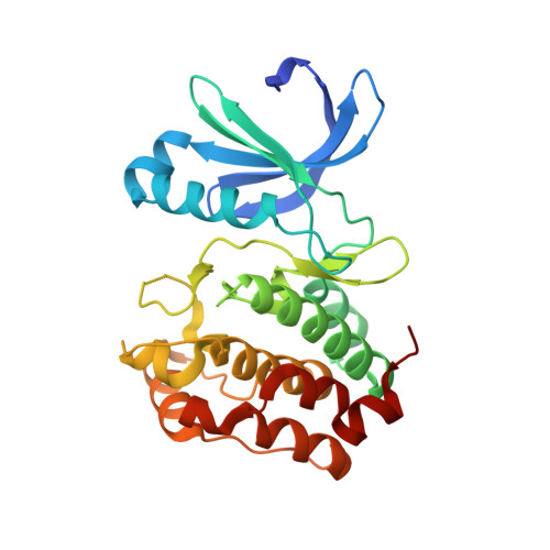
Computationally-guided optimization of small-molecule inhibitors of the Aurora A kinase-TPX2 protein-protein interaction.

Cole, D.J.Janecek, M.Stokes, J.E.Rossmann, M.Faver, J.C.McKenzie, G.J.Venkitaraman, A.R.Hyvonen, M.Spring, D.R.Huggins, D.J.Jorgensen, W.L.

(2017) Chem Commun (Camb) 53: 9372-9375

  • DOI: https://doi.org/10.1039/c7cc05379g
  • Primary Citation Related Structures: 
    5OBJ, 5OBR

  • PubMed Abstract: 

    Free energy perturbation theory, in combination with enhanced sampling of protein-ligand binding modes, is evaluated in the context of fragment-based drug design, and used to design two new small-molecule inhibitors of the Aurora A kinase-TPX2 protein-protein interaction.


  • Organizational Affiliation
    • Department of Chemistry, Yale University, New Haven, Connecticut 06520-8107, USA and School of Chemistry, Newcastle University, Newcastle upon Tyne NE1 7RU, UK. daniel.cole@ncl.ac.uk.

Macromolecule Content 

  • Total Structure Weight: 32.38 kDa 
  • Atom Count: 2,186 
  • Modeled Residue Count: 260 
  • Deposited Residue Count: 272 
  • Unique protein chains: 1

Macromolecules

Find similar proteins by:|  3D Structure
Entity ID: 1
MoleculeChains  Sequence LengthOrganismDetailsImage
Aurora kinase A272Homo sapiensMutation(s): 0 
Gene Names: AURKAAIKAIRK1ARK1AURAAYK1BTAKIAK1STK15STK6
EC: 2.7.11.1
UniProt & NIH Common Fund Data Resources
Find proteins for O14965 (Homo sapiens)
Explore O14965 
Go to UniProtKB:  O14965
PHAROS:  O14965
GTEx:  ENSG00000087586 
Entity Groups
Sequence Clusters30% Identity50% Identity70% Identity90% Identity95% Identity100% Identity
UniProt GroupO14965
Sequence Annotations
Expand
Reference Sequence

Small Molecules

Experimental Data & Validation

Experimental Data

  • Method: X-RAY DIFFRACTION
  • Resolution: 2.90 Å
  • R-Value Free:  0.245 (Depositor), 0.250 (DCC) 
  • R-Value Work:  0.214 (Depositor), 0.213 (DCC) 
  • R-Value Observed: 0.215 (Depositor) 
Space Group: P 61 2 2
Unit Cell:
Length ( Å )Angle ( ˚ )
a = 83.41α = 90
b = 83.41β = 90
c = 172.09γ = 120
Software Package:
Software NamePurpose
PHENIXrefinement
XDSdata reduction
XDSdata scaling
PHASERphasing

Structure Validation

View Full Validation Report


Ligand Structure Quality Assessment 


Entry History 

& Funding Information

Deposition Data


Funding OrganizationLocationGrant Number
Wellcome TrustUnited Kingdom090340/Z/09/Z

Revision History  (Full details and data files)

  • Version 1.0: 2017-08-09
    Type: Initial release
  • Version 1.1: 2017-08-30
    Changes: Database references
  • Version 1.2: 2024-05-08
    Changes: Data collection, Database references