5OBC | pdb_00005obc

X-ray structure of the adduct formed upon reaction of ribonuclease A with the compound fac-[RuII(CO)3Cl2(N3-IM), IM=imidazole


Experimental Data Snapshot

  • Method: X-RAY DIFFRACTION
  • Resolution: 2.07 Å
  • R-Value Free: 
    0.272 (Depositor), 0.272 (DCC) 
  • R-Value Work: 
    0.181 (Depositor), 0.189 (DCC) 
  • R-Value Observed: 
    0.186 (Depositor) 

Starting Model: experimental
View more details

wwPDB Validation 3D Report Full Report

Validation slider image for 5OBC

This is version 1.3 of the entry. See complete history

Literature

Ru-Based CO releasing molecules with azole ligands: interaction with proteins and the CO release mechanism disclosed by X-ray crystallography.

Pontillo, N.Ferraro, G.Messori, L.Tamasi, G.Merlino, A.

(2017) Dalton Trans 46: 9621-9629

  • DOI: https://doi.org/10.1039/c7dt01991b
  • Primary Citation Related Structures: 
    5OB6, 5OB7, 5OB8, 5OB9, 5OBC, 5OBD, 5OBE

  • PubMed Abstract: 

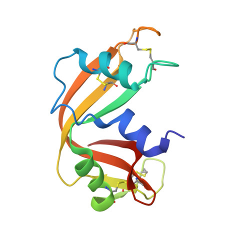
    fac-[Ru II (CO) 3 Cl 2 (N 3 -Imidazole)] (Ru II IM), fac-[Ru II (CO) 3 Cl 2 (N 3 -methyl-imidazole)] (Ru II MIM) and fac-[Ru II (CO) 3 Cl 2 (N 3 -methyl-benzimidazole)] (Ru II MBI) are three ruthenium based CO releasing molecules (Ru-CORMs) that are cytotoxic towards ovarian and colon carcinoma cell lines. Detailed structural information on the adducts formed upon reaction of Ru II IM and Ru II MIM with hen egg white lysozyme and of the three Ru-CORMs with bovine pancreatic ribonuclease is provided here by X-ray crystallography. Comparative analysis of seven crystal structures of these adducts allows one to delineate some general trends in the reactivity of these Ru-CORMs with proteins. Indeed, in all cases Ru-CORMs bind these model systems upon detachment of the azole ligand and concomitant coordination to a protein His or Asp residue. Apparently the three Ru-CORMs progressively dissociate losing azoles, chlorides, and one or two CO molecules. Data were compared with those reported in the literature for adducts of the same proteins with other Ru-CORMs and with in-solution data previously obtained on the same systems. These results are potentially useful for a better understanding of the chemistry, potential toxicity and mechanism of actions of these interesting Ru-CORMs and are helpful in defining the molecular mechanisms of CO release.


  • Organizational Affiliation
    • Department of Chemical Sciences, University of Naples Federico II, Complesso Universitario di Monte Sant' Angelo, Via Cinthia, I-80126, Napoli, Italy. antonello.merlino@unina.it.

Macromolecule Content 

  • Total Structure Weight: 28.28 kDa 
  • Atom Count: 2,139 
  • Modeled Residue Count: 248 
  • Deposited Residue Count: 248 
  • Unique protein chains: 1

Macromolecules

Find similar proteins by:|  3D Structure
Entity ID: 1
MoleculeChains  Sequence LengthOrganismDetailsImage
Ribonuclease pancreatic
A, B
124Bos taurusMutation(s): 0 
EC: 3.1.27.5 (PDB Primary Data), 4.6.1.18 (UniProt)
UniProt
Find proteins for P61823 (Bos taurus)
Explore P61823 
Go to UniProtKB:  P61823
Entity Groups
Sequence Clusters30% Identity50% Identity70% Identity90% Identity95% Identity100% Identity
UniProt GroupP61823
Sequence Annotations
Expand
Reference Sequence

Experimental Data & Validation

Experimental Data

  • Method: X-RAY DIFFRACTION
  • Resolution: 2.07 Å
  • R-Value Free:  0.272 (Depositor), 0.272 (DCC) 
  • R-Value Work:  0.181 (Depositor), 0.189 (DCC) 
  • R-Value Observed: 0.186 (Depositor) 
Space Group: C 1 2 1
Unit Cell:
Length ( Å )Angle ( ˚ )
a = 100.552α = 90
b = 32.698β = 90.64
c = 74.688γ = 90
Software Package:
Software NamePurpose
REFMACrefinement
HKL-2000data reduction
HKL-2000data scaling
PHASERphasing

Structure Validation

View Full Validation Report


Entry History 

Deposition Data

Revision History  (Full details and data files)

  • Version 1.0: 2017-07-26
    Type: Initial release
  • Version 1.1: 2017-08-02
    Changes: Database references
  • Version 1.2: 2024-01-17
    Changes: Advisory, Data collection, Database references, Refinement description
  • Version 1.3: 2024-11-06
    Changes: Structure summary