5OAY | pdb_00005oay

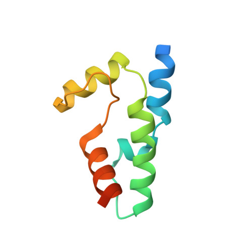
M. tuberculosis [4Fe-4S] protein WhiB1 is a four-helix bundle that forms a NO-sensitive complex with sigmaA and regulates the major virulence factor ESX-1


Experimental Data Snapshot

  • Method: SOLUTION NMR
  • Conformers Calculated: 62 
  • Conformers Submitted: 10 
  • Selection Criteria: structures with the lowest energy 

wwPDB Validation 3D Report Full Report

Validation slider image for 5OAY

This is version 2.2 of the entry. See complete history. 

Literature

Structure of a Wbl protein and implications for NO sensing by M. tuberculosis.

Kudhair, B.K., Hounslow, A.M., Rolfe, M.D., Crack, J.C., Hunt, D.M., Buxton, R.S., Smith, L.J., Le Brun, N.E., Williamson, M.P., Green, J.

(2017) Nat Commun 8: 2280-2280

  • DOI: https://doi.org/10.1038/s41467-017-02418-y
  • Primary Citation Related Structures: 
    5OAY

  • PubMed Abstract: 

    Mycobacterium tuberculosis causes pulmonary tuberculosis (TB) and claims ~1.8 million human lives per annum. Host nitric oxide (NO) is important in controlling TB infection. M. tuberculosis WhiB1 is a NO-responsive Wbl protein (actinobacterial iron-sulfur proteins first identified in the 1970s). Until now, the structure of a Wbl protein has not been available. Here a NMR structural model of WhiB1 reveals that Wbl proteins are four-helix bundles with a core of three α-helices held together by a [4Fe-4S] cluster. The iron-sulfur cluster is required for formation of a complex with the major sigma factor (σ A ) and reaction with NO disassembles this complex. The WhiB1 structure suggests that loss of the iron-sulfur cluster (by nitrosylation) permits positively charged residues in the C-terminal helix to engage in DNA binding, triggering a major reprogramming of gene expression that includes components of the virulence-critical ESX-1 secretion system.


  • Organizational Affiliation: 
    • Molecular Biology and Biotechnology, University of Sheffield, Sheffield, S10 2TN, UK.

Macromolecule Content 

  • Total Structure Weight: 10.74 kDa 
  • Atom Count: 597 
  • Modeled Residue Count: 76 
  • Deposited Residue Count: 94 
  • Unique protein chains: 1

Macromolecules

Find similar proteins by:|  3D Structure
Entity ID: 1
MoleculeChains  Sequence LengthOrganismDetailsImage
Transcriptional regulator WhiB194Mycobacterium tuberculosis H37RvMutation(s): 0 
Gene Names: whiB1, Rv3219
UniProt
Find proteins for P9WF43 (Mycobacterium tuberculosis (strain ATCC 25618 / H37Rv))
Explore P9WF43 
Go to UniProtKB:  P9WF43
Entity Groups
Sequence Clusters30% Identity50% Identity70% Identity90% Identity95% Identity100% Identity
UniProt GroupP9WF43
Sequence Annotations
Expand
Reference Sequence

Small Molecules

Ligands 1 Unique
IDChains Name / Formula / InChI Key2D Diagram3D Interactions
SF4
(Subject of Investigation/LOI)

Query on SF4


Download:Ideal Coordinates CCD File
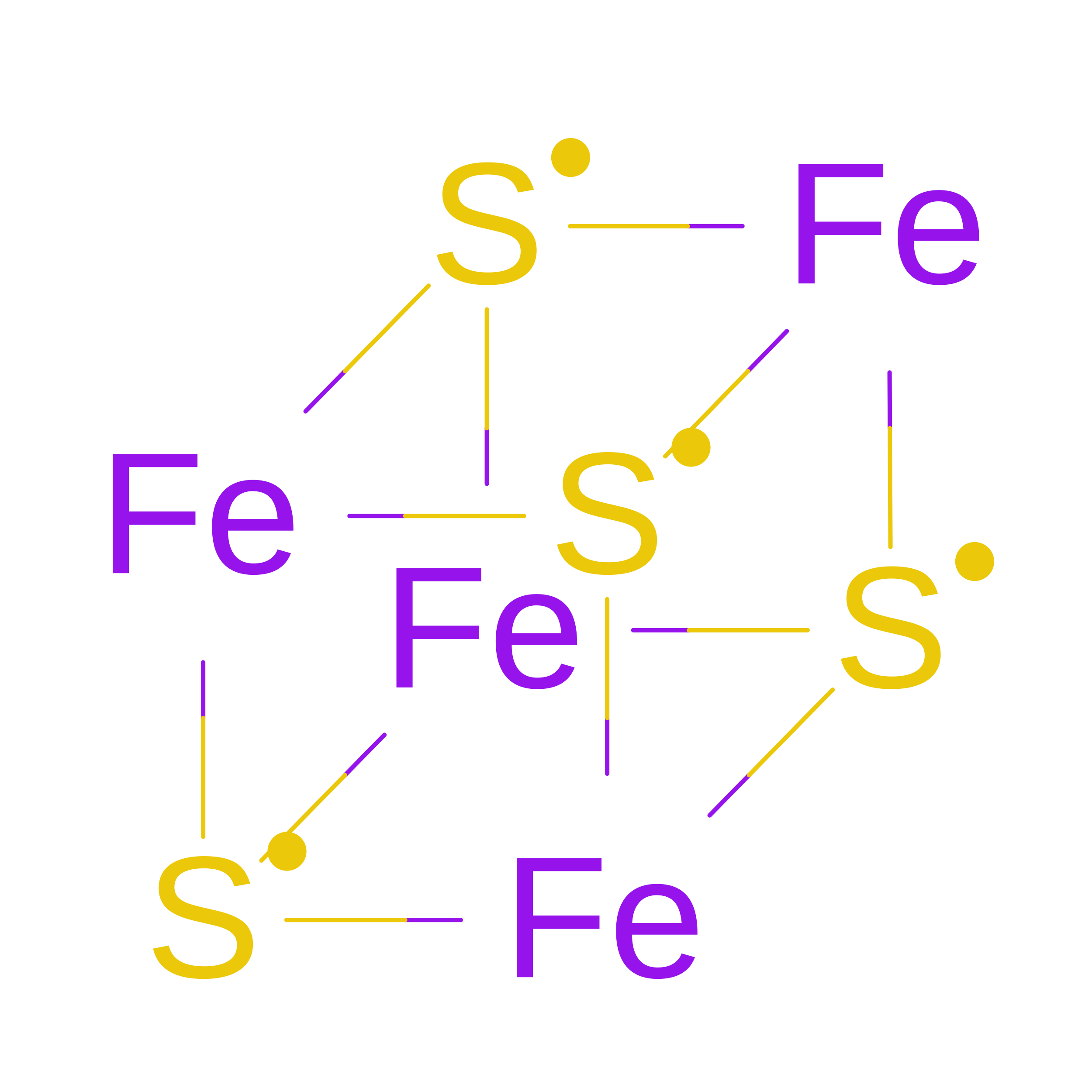
B [auth A]IRON/SULFUR CLUSTER
Fe4 S4
LJBDFODJNLIPKO-UHFFFAOYSA-N

Experimental Data & Validation

Experimental Data

  • Method: SOLUTION NMR
  • Conformers Calculated: 62 
  • Conformers Submitted: 10 
  • Selection Criteria: structures with the lowest energy 

Structure Validation

View Full Validation Report


Entry History 

& Funding Information

Deposition Data


Funding OrganizationLocationGrant Number
Biotechnology and Biological Sciences Research CouncilUnited KingdomBB/K000071/1
Biotechnology and Biological Sciences Research CouncilUnited KingdomBB/L008114/1

Revision History  (Full details and data files)

  • Version 1.0: 2018-01-03
    Type: Initial release
  • Version 2.0: 2018-01-10
    Changes: Atomic model
  • Version 2.1: 2024-05-15
    Changes: Data collection, Database references
  • Version 2.2: 2026-08-26
    Type: Remediation
    Reason: Metalloprotein remediation
    Changes: Derived calculations, Structure summary