5O9X | pdb_00005o9x

Crystal structure of Aspergillus fumigatus N-acetylphosphoglucosamine mutate S69A in complex with glucose1,6bisphosphate


Experimental Data Snapshot

  • Method: X-RAY DIFFRACTION
  • Resolution: 1.90 Å
  • R-Value Free: 
    0.201 (Depositor), 0.200 (DCC) 
  • R-Value Work: 
    0.165 (Depositor), 0.166 (DCC) 
  • R-Value Observed: 
    0.165 (Depositor) 

wwPDB Validation 3D Report Full Report

Validation slider image for 5O9X

Ligand Structure Quality Assessment 


This is version 1.3 of the entry. See complete history

Literature

Evidence for substrate-assisted catalysis inN-acetylphosphoglucosamine mutase.

Raimi, O.G.Hurtado-Guerrero, R.van Aalten, D.M.F.

(2018) Biochem J 475: 2547-2557

  • DOI: https://doi.org/10.1042/BCJ20180172
  • Primary Citation Related Structures: 
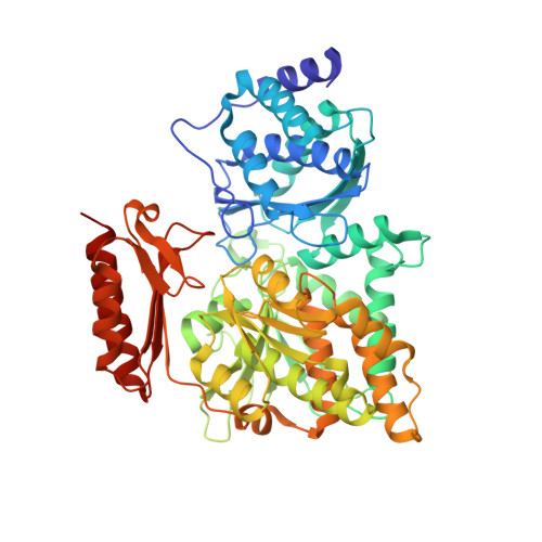
    5O9X, 5OAW

  • PubMed Abstract: 

    N -acetylphosphoglucosamine mutase (AGM1) is a key component of the hexosamine biosynthetic pathway that produces UDP-GlcNAc, an essential precursor for a wide range of glycans in eukaryotes. AGM belongs to the α-d-phosphohexomutase metalloenzyme superfamily and catalyzes the interconversion of N -acetylglucosamine-6-phosphate (GlcNAc-6P) to N -acetylglucosamine-1-phosphate (GlcNAc-1P) through N -acetylglucosamine-1,6-bisphosphate (GlcNAc-1,6-bisP) as the catalytic intermediate. Although there is an understanding of the phosphoserine-dependent catalytic mechanism at enzymatic and structural level, the identity of the requisite catalytic base in AGM1/phosphoglucomutases is as yet unknown. Here, we present crystal structures of a Michaelis complex of AGM1 with GlcNAc-6P and Mg 2+ , and a complex of the inactive Ser69Ala mutant together with glucose-1,6-bisphosphate (Glc-1,6-bisP) that represents key snapshots along the reaction co-ordinate. Together with mutagenesis, these structures reveal that the phosphate group of the hexose-1,6-bisP intermediate may act as the catalytic base.


  • Organizational Affiliation
    • Division of Gene Regulation and Expression, School of Life Sciences, University of Dundee, Dundee DD1 5EH, U.K.

Macromolecule Content 

  • Total Structure Weight: 60.51 kDa 
  • Atom Count: 4,657 
  • Modeled Residue Count: 541 
  • Deposited Residue Count: 549 
  • Unique protein chains: 1

Macromolecules

Find similar proteins by:|  3D Structure
Entity ID: 1
MoleculeChains  Sequence LengthOrganismDetailsImage
Phosphoacetylglucosamine mutase549Aspergillus lentulusMutation(s): 1 
Gene Names: ALT_8497
EC: 5.4.2.3

Experimental Data & Validation

Experimental Data

  • Method: X-RAY DIFFRACTION
  • Resolution: 1.90 Å
  • R-Value Free:  0.201 (Depositor), 0.200 (DCC) 
  • R-Value Work:  0.165 (Depositor), 0.166 (DCC) 
  • R-Value Observed: 0.165 (Depositor) 
Space Group: P 21 21 21
Unit Cell:
Length ( Å )Angle ( ˚ )
a = 80.86α = 90
b = 87.574β = 90
c = 91.867γ = 90
Software Package:
Software NamePurpose
REFMACrefinement
XDSdata reduction
SCALAdata scaling
MOLREPphasing

Structure Validation

View Full Validation Report


Ligand Structure Quality Assessment 


Entry History 

Deposition Data

Revision History  (Full details and data files)

  • Version 1.0: 2018-07-11
    Type: Initial release
  • Version 1.1: 2018-08-29
    Changes: Data collection, Database references
  • Version 1.2: 2020-07-29
    Type: Remediation
    Reason: Carbohydrate remediation
    Changes: Data collection, Derived calculations, Structure summary
  • Version 1.3: 2024-05-08
    Changes: Data collection, Database references, Structure summary