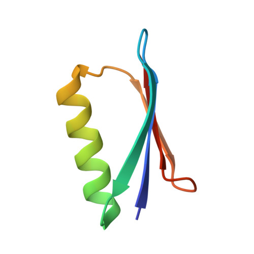
5O94

X-ray structure of a zinc binding GB1 mutant


Experimental Data Snapshot

  • Method: X-RAY DIFFRACTION
  • Resolution: 1.90 Å
  • R-Value Free: 0.234 
  • R-Value Work: 0.206 
  • R-Value Observed: 0.208 

Starting Model: experimental
View more details

wwPDB Validation   3D Report Full Report


This is version 1.2 of the entry. See complete history


Literature

Genetic Algorithm Based Design and Experimental Characterization of a Highly Thermostable Metalloprotein.

Bozkurt, E.Perez, M.A.S.Hovius, R.Browning, N.J.Rothlisberger, U.

(2018) J Am Chem Soc 140: 4517-4521

  • DOI: https://doi.org/10.1021/jacs.7b10660
  • Primary Citation of Related Structures:  
    5O94, 5OFS

  • PubMed Abstract: 

    The development of thermostable and solvent-tolerant metalloproteins is a long-sought goal for many applications in synthetic biology and biotechnology. In this work, we were able to engineer a highly thermostable and organic solvent-stable metallo variant of the B1 domain of protein G (GB1) with a tetrahedral zinc binding site reminiscent of the one of thermolysin. Promising candidates were designed computationally by applying a protocol based on classical and first-principles molecular dynamics simulations in combination with genetic algorithm optimization. The most promising of the computationally predicted mutants was expressed and structurally characterized and yielded a highly thermostable protein. The experimental results thus confirm the predictive power of the applied computational protein engineering approach for the de novo design of highly stable metalloproteins.


  • Organizational Affiliation

    Laboratory of Computational Chemistry and Biochemistry , École Polytechnique Fédérale de Lausanne , CH-1015 Lausanne , Switzerland.


Macromolecules
Find similar proteins by:  (by identity cutoff)  |  3D Structure
Entity ID: 1
MoleculeChains Sequence LengthOrganismDetailsImage
Metal binding GB156Streptococcus sp.Mutation(s): 0 
Entity Groups  
Sequence Clusters30% Identity50% Identity70% Identity90% Identity95% Identity100% Identity
Sequence Annotations
Expand
  • Reference Sequence
Small Molecules
Ligands 6 Unique
IDChains Name / Formula / InChI Key2D Diagram3D Interactions
MPD
Query on MPD

Download Ideal Coordinates CCD File 
C [auth A](4S)-2-METHYL-2,4-PENTANEDIOL
C6 H14 O2
SVTBMSDMJJWYQN-YFKPBYRVSA-N
SO4
Query on SO4

Download Ideal Coordinates CCD File 
E [auth A],
F [auth A],
G [auth A]
SULFATE ION
O4 S
QAOWNCQODCNURD-UHFFFAOYSA-L
GOL
Query on GOL

Download Ideal Coordinates CCD File 
K [auth A],
L [auth A]
GLYCEROL
C3 H8 O3
PEDCQBHIVMGVHV-UHFFFAOYSA-N
ZN (Subject of Investigation/LOI)
Query on ZN

Download Ideal Coordinates CCD File 
B [auth A]ZINC ION
Zn
PTFCDOFLOPIGGS-UHFFFAOYSA-N
EDO
Query on EDO

Download Ideal Coordinates CCD File 
H [auth A],
I [auth A],
J [auth A]
1,2-ETHANEDIOL
C2 H6 O2
LYCAIKOWRPUZTN-UHFFFAOYSA-N
NA
Query on NA

Download Ideal Coordinates CCD File 
D [auth A]SODIUM ION
Na
FKNQFGJONOIPTF-UHFFFAOYSA-N
Experimental Data & Validation

Experimental Data

  • Method: X-RAY DIFFRACTION
  • Resolution: 1.90 Å
  • R-Value Free: 0.234 
  • R-Value Work: 0.206 
  • R-Value Observed: 0.208 
  • Space Group: P 32 2 1
Unit Cell:
Length ( Å )Angle ( ˚ )
a = 54.178α = 90
b = 54.178β = 90
c = 81.077γ = 120
Software Package:
Software NamePurpose
REFMACrefinement
XDSdata reduction
XDSdata scaling
PHASERphasing

Structure Validation

View Full Validation Report


Entry History & Funding Information

Deposition Data


Funding OrganizationLocationGrant Number
Swiss National Science FoundationSwitzerland200020-146645

Revision History  (Full details and data files)

  • Version 1.0: 2018-04-04
    Type: Initial release
  • Version 1.1: 2018-04-11
    Changes: Data collection, Database references, Derived calculations, Structure summary
  • Version 1.2: 2024-01-17
    Changes: Data collection, Database references, Derived calculations, Refinement description, Structure summary