5O7Y | pdb_00005o7y

Thebaine 6-O-demethylase (T6ODM) from Papaver somniferum in complex with succinate


Experimental Data Snapshot

  • Method: X-RAY DIFFRACTION
  • Resolution: 1.97 Å
  • R-Value Free: 
    0.207 (Depositor), 0.215 (DCC) 
  • R-Value Work: 
    0.163 (Depositor), 0.172 (DCC) 
  • R-Value Observed: 
    0.165 (Depositor) 

Starting Model: experimental
View more details

wwPDB Validation 3D Report Full Report

Validation slider image for 5O7Y

Ligand Structure Quality Assessment 


This is version 1.4 of the entry. See complete history

Literature

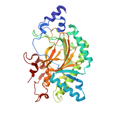
Crystal structure of thebaine 6-O-demethylase from the morphine biosynthesis pathway.

Kluza, A.Niedzialkowska, E.Kurpiewska, K.Wojdyla, Z.Quesne, M.Kot, E.Porebski, P.J.Borowski, T.

(2018) J Struct Biol 202: 229-235

  • DOI: https://doi.org/10.1016/j.jsb.2018.01.007
  • Primary Citation Related Structures: 
    5O7Y, 5O9W

  • PubMed Abstract: 

    Thebaine 6-O-demethylase (T6ODM) from Papaver somniferum (opium poppy), which belongs to the non-heme 2-oxoglutarate/Fe(II)-dependent dioxygenases (ODD) family, is a key enzyme in the morphine biosynthesis pathway. Initially, T6ODM was characterized as an enzyme catalyzing O-demethylation of thebaine to neopinone and oripavine to morphinone. However, the substrate range of T6ODM was recently expanded to a number of various benzylisoquinoline alkaloids. Here, we present crystal structures of T6ODM in complexes with 2-oxoglutarate (T6ODM:2OG, PDB: 5O9W) and succinate (T6ODM:SIN, PDB: 5O7Y). Both metal and 2OG binding sites display similarity to other proteins from the ODD family, but T6ODM is characterized by an exceptionally large substrate binding cavity, whose volume can partially explain the promiscuity of this enzyme. Moreover, the size of the cavity allows for binding of multiple molecules at once, posing a question about the substrate-driven specificity of the enzyme.


  • Organizational Affiliation
    • Jerzy Haber Institute of Catalysis and Surface Chemistry, Polish Academy of Sciences, Niezapominajek 8, PL-30239 Krakow, Poland.

Macromolecule Content 

  • Total Structure Weight: 42.38 kDa 
  • Atom Count: 3,270 
  • Modeled Residue Count: 358 
  • Deposited Residue Count: 367 
  • Unique protein chains: 1

Macromolecules

Find similar proteins by:|  3D Structure
Entity ID: 1
MoleculeChains  Sequence LengthOrganismDetailsImage
Thebaine 6-O-demethylase367Papaver somniferumMutation(s): 0 
Gene Names: T6ODMDIOX1
EC: 1.14.11.31 (PDB Primary Data), 2.1.1 (UniProt)
UniProt
Find proteins for D4N500 (Papaver somniferum)
Explore D4N500 
Go to UniProtKB:  D4N500
Entity Groups
Sequence Clusters30% Identity50% Identity70% Identity90% Identity95% Identity100% Identity
UniProt GroupD4N500
Sequence Annotations
Expand
Reference Sequence

Small Molecules

Ligands 4 Unique
IDChains Name / Formula / InChI Key2D Diagram3D Interactions
SIN
(Subject of Investigation/LOI)

Query on SIN


Download:Ideal Coordinates CCD File
C [auth A]SUCCINIC ACID
C4 H6 O4
KDYFGRWQOYBRFD-UHFFFAOYSA-N
PEG

Query on PEG


Download:Ideal Coordinates CCD File
U [auth A],
V [auth A]
DI(HYDROXYETHYL)ETHER
C4 H10 O3
MTHSVFCYNBDYFN-UHFFFAOYSA-N
EDO

Query on EDO


Download:Ideal Coordinates CCD File
D [auth A]
E [auth A]
F [auth A]
G [auth A]
H [auth A]
D [auth A],
E [auth A],
F [auth A],
G [auth A],
H [auth A],
I [auth A],
J [auth A],
K [auth A],
L [auth A],
M [auth A],
N [auth A],
O [auth A],
P [auth A],
Q [auth A],
R [auth A],
S [auth A],
T [auth A]
1,2-ETHANEDIOL
C2 H6 O2
LYCAIKOWRPUZTN-UHFFFAOYSA-N
NI
(Subject of Investigation/LOI)

Query on NI


Download:Ideal Coordinates CCD File
B [auth A]NICKEL (II) ION
Ni
VEQPNABPJHWNSG-UHFFFAOYSA-N

Experimental Data & Validation

Experimental Data

  • Method: X-RAY DIFFRACTION
  • Resolution: 1.97 Å
  • R-Value Free:  0.207 (Depositor), 0.215 (DCC) 
  • R-Value Work:  0.163 (Depositor), 0.172 (DCC) 
  • R-Value Observed: 0.165 (Depositor) 
Space Group: P 21 21 21
Unit Cell:
Length ( Å )Angle ( ˚ )
a = 47.231α = 90
b = 87.028β = 90
c = 94.171γ = 90
Software Package:
Software NamePurpose
REFMACrefinement
HKL-3000data reduction
HKL-3000data scaling
HKL-3000phasing
MOLREPphasing

Structure Validation

View Full Validation Report


Ligand Structure Quality Assessment 


Entry History 

& Funding Information

Deposition Data


Funding OrganizationLocationGrant Number
National Science Centre, PolandPolandUMO-2014/14/E/NZ1/00053

Revision History  (Full details and data files)

  • Version 1.0: 2018-02-14
    Type: Initial release
  • Version 1.1: 2018-04-04
    Changes: Data collection
  • Version 1.2: 2018-05-23
    Changes: Data collection, Database references
  • Version 1.3: 2020-04-22
    Changes: Database references
  • Version 1.4: 2024-01-17
    Changes: Data collection, Database references, Derived calculations, Refinement description