5O6S | pdb_00005o6s

UbV.B4R, a dimeric ubiquitin variant binding to BIRC4 RING


Experimental Data Snapshot

  • Method: X-RAY DIFFRACTION
  • Resolution: 2.90 Å
  • R-Value Free: 
    0.309 (Depositor) 
  • R-Value Work: 
    0.261 (Depositor) 
  • R-Value Observed: 
    0.263 (Depositor) 

Starting Model: experimental
View more details

wwPDB Validation 3D Report Full Report

Currently 5O6S does not have a validation slider image.


This is version 1.1 of the entry. See complete history

Literature

A General Strategy for Discovery of Inhibitors and Activators of RING and U-box E3 Ligases with Ubiquitin Variants.

Gabrielsen, M.Buetow, L.Nakasone, M.A.Ahmed, S.F.Sibbet, G.J.Smith, B.O.Zhang, W.Sidhu, S.S.Huang, D.T.

(2017) Mol Cell 68: 456-470.e10

  • DOI: https://doi.org/10.1016/j.molcel.2017.09.027
  • Primary Citation Related Structures: 
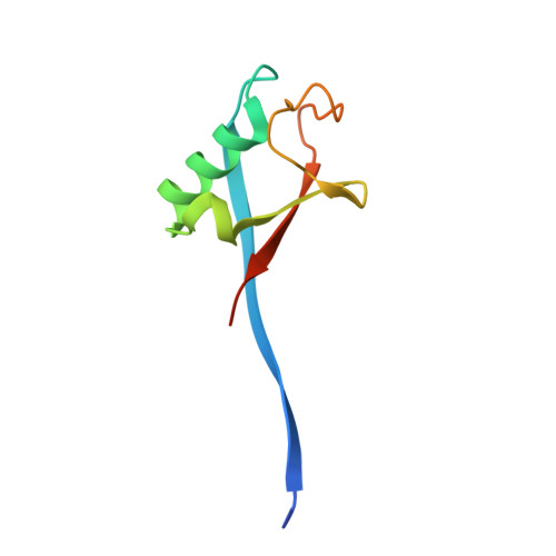
    5O6S, 5O6T, 5O75, 5O76

  • PubMed Abstract: 

    RING and U-box E3 ubiquitin ligases regulate diverse eukaryotic processes and have been implicated in numerous diseases, but targeting these enzymes remains a major challenge. We report the development of three ubiquitin variants (UbVs), each binding selectively to the RING or U-box domain of a distinct E3 ligase: monomeric UBE4B, phosphorylated active CBL, or dimeric XIAP. Structural and biochemical analyses revealed that UbVs specifically inhibited the activity of UBE4B or phosphorylated CBL by blocking the E2∼Ub binding site. Surprisingly, the UbV selective for dimeric XIAP formed a dimer to stimulate E3 activity by stabilizing the closed E2∼Ub conformation. We further verified the inhibitory and stimulatory functions of UbVs in cells. Our work provides a general strategy to inhibit or activate RING/U-box E3 ligases and provides a resource for the research community to modulate these enzymes.


  • Organizational Affiliation
    • Cancer Research UK Beatson Institute, Garscube Estate, Switchback Road, Glasgow G61 1BD, UK.

Macromolecule Content 

  • Total Structure Weight: 55.45 kDa 
  • Atom Count: 3,613 
  • Modeled Residue Count: 450 
  • Deposited Residue Count: 498 
  • Unique protein chains: 1

Macromolecules

Find similar proteins by:|  3D Structure
Entity ID: 1
MoleculeChains  Sequence LengthOrganismDetailsImage
Polyubiquitin-B
A, B, C, D, E
A, B, C, D, E, F
83Homo sapiensMutation(s): 0 
Gene Names: UBB

Experimental Data & Validation

Experimental Data

  • Method: X-RAY DIFFRACTION
  • Resolution: 2.90 Å
  • R-Value Free:  0.309 (Depositor) 
  • R-Value Work:  0.261 (Depositor) 
  • R-Value Observed: 0.263 (Depositor) 
Space Group: I 1 2 1
Unit Cell:
Length ( Å )Angle ( ˚ )
a = 48.213α = 90
b = 71.669β = 94.97
c = 118.033γ = 90
Software Package:
Software NamePurpose
BUSTERrefinement
DIALSdata reduction
Aimlessdata scaling
PHASERphasing

Structure Validation

View Full Validation Report

Currently 5O6S does not have a validation slider image.


Entry History 

& Funding Information

Deposition Data


Funding OrganizationLocationGrant Number
Cancer Research UKUnited KingdomC596/A23278
European Research CouncilUnited Kingdom647849

Revision History  (Full details and data files)

  • Version 1.0: 2017-11-01
    Type: Initial release
  • Version 1.1: 2024-01-17
    Changes: Advisory, Data collection, Database references, Refinement description