5O56 | pdb_00005o56

Glycogen Phosphorylase b in complex with 29b


Experimental Data Snapshot

  • Method: X-RAY DIFFRACTION
  • Resolution: 2.45 Å
  • R-Value Free: 
    0.229 (Depositor), 0.229 (DCC) 
  • R-Value Work: 
    0.164 (Depositor), 0.165 (DCC) 
  • R-Value Observed: 
    0.168 (Depositor) 

wwPDB Validation 3D Report Full Report

Validation slider image for 5O56

Ligand Structure Quality Assessment 


This is version 1.2 of the entry. See complete history

Literature

Nanomolar Inhibitors of Glycogen Phosphorylase Based on beta-d-Glucosaminyl Heterocycles: A Combined Synthetic, Enzyme Kinetic, and Protein Crystallography Study.

Bokor, E.Kyriakis, E.Solovou, T.G.A.Koppany, C.Kantsadi, A.L.Szabo, K.E.Szakacs, A.Stravodimos, G.A.Docsa, T.Skamnaki, V.T.Zographos, S.E.Gergely, P.Leonidas, D.D.Somsak, L.

(2017) J Med Chem 60: 9251-9262

  • DOI: https://doi.org/10.1021/acs.jmedchem.7b01056
  • Primary Citation Related Structures: 
    5O50, 5O52, 5O54, 5O56

  • PubMed Abstract: 

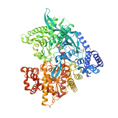
    Aryl substituted 1-(β-d-glucosaminyl)-1,2,3-triazoles as well as C-β-d-glucosaminyl 1,2,4-triazoles and imidazoles were synthesized and tested as inhibitors against muscle and liver isoforms of glycogen phosphorylase (GP). While the N-β-d-glucosaminyl 1,2,3-triazoles showed weak or no inhibition, the C-β-d-glucosaminyl derivatives had potent activity, and the best inhibitor was the 2-(β-d-glucosaminyl)-4(5)-(2-naphthyl)-imidazole with a K i value of 143 nM against human liver GPa. An X-ray crystallography study of the rabbit muscle GPb inhibitor complexes revealed structural features of the strong binding and offered an explanation for the differences in inhibitory potency between glucosyl and glucosaminyl derivatives and also for the differences between imidazole and 1,2,4-triazole analogues.


  • Organizational Affiliation
    • Department of Organic Chemistry, University of Debrecen , POB 400, H-4002 Debrecen, Hungary.

Macromolecule Content 

  • Total Structure Weight: 98.03 kDa 
  • Atom Count: 6,851 
  • Modeled Residue Count: 810 
  • Deposited Residue Count: 843 
  • Unique protein chains: 1

Macromolecules

Find similar proteins by:|  3D Structure
Entity ID: 1
MoleculeChains  Sequence LengthOrganismDetailsImage
Glycogen phosphorylase, muscle form843Oryctolagus cuniculusMutation(s): 0 
EC: 2.4.1.1
UniProt
Find proteins for P00489 (Oryctolagus cuniculus)
Explore P00489 
Go to UniProtKB:  P00489
Entity Groups
Sequence Clusters30% Identity50% Identity70% Identity90% Identity95% Identity100% Identity
UniProt GroupP00489
Sequence Annotations
Expand
Reference Sequence

Small Molecules

Ligands 2 Unique
IDChains Name / Formula / InChI Key2D Diagram3D Interactions
9L8

Query on 9L8


Download:Ideal Coordinates CCD File
C [auth A](2~{R},3~{S},4~{R},5~{R},6~{R})-5-azanyl-2-(hydroxymethyl)-6-(3-naphthalen-2-yl-1~{H}-1,2,4-triazol-5-yl)oxane-3,4-diol
C18 H20 N4 O4
IMNCDLXOKIGZHY-OXGONZEZSA-N
PLP

Query on PLP


Download:Ideal Coordinates CCD File
B [auth A]PYRIDOXAL-5'-PHOSPHATE
C8 H10 N O6 P
NGVDGCNFYWLIFO-UHFFFAOYSA-N

Experimental Data & Validation

Experimental Data

  • Method: X-RAY DIFFRACTION
  • Resolution: 2.45 Å
  • R-Value Free:  0.229 (Depositor), 0.229 (DCC) 
  • R-Value Work:  0.164 (Depositor), 0.165 (DCC) 
  • R-Value Observed: 0.168 (Depositor) 
Space Group: P 43 21 2
Unit Cell:
Length ( Å )Angle ( ˚ )
a = 128.548α = 90
b = 128.548β = 90
c = 116.141γ = 90
Software Package:
Software NamePurpose
REFMACrefinement
CrysalisProdata reduction
Aimlessdata scaling

Structure Validation

View Full Validation Report


Ligand Structure Quality Assessment 


Entry History 

Deposition Data

Revision History  (Full details and data files)

  • Version 1.0: 2017-09-27
    Type: Initial release
  • Version 1.1: 2017-11-29
    Changes: Database references
  • Version 1.2: 2025-04-09
    Changes: Data collection, Database references, Structure summary