5O4Q | pdb_00005o4q

Crystal Structure of mutant M54L/M64L/M96L of Two-Domain Laccase from Streptomyces griseoflavus with 0.25 mM copper sulfate on growth medium


Experimental Data Snapshot

  • Method: X-RAY DIFFRACTION
  • Resolution: 1.90 Å
  • R-Value Free: 
    0.213 (Depositor), 0.212 (DCC) 
  • R-Value Work: 
    0.187 (Depositor), 0.189 (DCC) 
  • R-Value Observed: 
    0.188 (Depositor) 

Starting Model: experimental
View more details

wwPDB Validation 3D Report Full Report

Validation slider image for 5O4Q

This is version 1.3 of the entry. See complete history

Literature

Incorporation of Copper Ions into T2/T3 Centers of Two-Domain Laccases.

Gabdulkhakov, A.G.Kostareva, O.S.Kolyadenko, I.A.Mikhaylina, A.O.Trubitsina, L.I.Tishchenko, S.V.

(2018) Mol Biol (Mosk) 52: 29-35

  • DOI: https://doi.org/10.7868/S0026898418010056
  • Primary Citation Related Structures: 
    5O3K, 5O4I, 5O4Q

  • PubMed Abstract: 

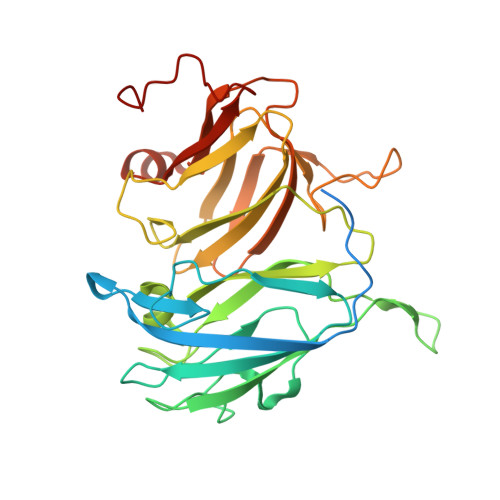
    Laccase belongs to the family of copper-containing oxidases. A study was made of the mechanism that sustains the incorporation of copper ions into the T2/T3 centers of recombinant two-domain laccase Streptomyces griseoflavus Ac-993. The occupancy of the T3 center by copper ions was found to increase with an increasing copper content in the culture medium and after dialysis of the protein preparation against a copper sulfate-containing buffer. The T2 center was filled only when overproducer strain cells were grown at a higher copper concentration in the medium. Two-domain laccases were assumed to possess a channel that serves to deliver copper ions to the T3 center during the formation of the three-dimensional laccase conformation and dialysis of the protein preparation. A narrower channel leads to the T2 center in two-domain laccases compared with three-domain ones, rendering the center less accessible for copper atoms. The incorporation of copper ions into the T2 center of two-domain laccases is likely to occur in the course of their biosynthesis or the formation of a functional trimer.


  • Organizational Affiliation
    • Institute of Protein Research, Russian Academy of Sciences, Pushchino, Moscow oblast, 142290 Russia.

Macromolecule Content 

  • Total Structure Weight: 418.54 kDa 
  • Atom Count: 26,408 
  • Modeled Residue Count: 3,329 
  • Deposited Residue Count: 3,864 
  • Unique protein chains: 1

Macromolecules

Find similar proteins by:|  3D Structure
Entity ID: 1
MoleculeChains  Sequence LengthOrganismDetailsImage
Two-domain laccase
A, B, C, D, E
A, B, C, D, E, F, G, H, I, J, K, L
322Streptomyces griseoflavusMutation(s): 3 
EC: 1.10.3.2
UniProt
Find proteins for A0A0M4FJ81 (Streptomyces griseoflavus)
Explore A0A0M4FJ81 
Go to UniProtKB:  A0A0M4FJ81
Entity Groups
Sequence Clusters30% Identity50% Identity70% Identity90% Identity95% Identity100% Identity
UniProt GroupA0A0M4FJ81
Sequence Annotations
Expand
Reference Sequence

Small Molecules

Ligands 6 Unique
IDChains Name / Formula / InChI Key2D Diagram3D Interactions
TRS

Query on TRS


Download:Ideal Coordinates CCD File
NA [auth J],
O [auth A]
2-AMINO-2-HYDROXYMETHYL-PROPANE-1,3-DIOL
C4 H12 N O3
LENZDBCJOHFCAS-UHFFFAOYSA-O
SO4

Query on SO4


Download:Ideal Coordinates CCD File
QA [auth K]SULFATE ION
O4 S
QAOWNCQODCNURD-UHFFFAOYSA-L
GOL

Query on GOL


Download:Ideal Coordinates CCD File
S [auth B]GLYCEROL
C3 H8 O3
PEDCQBHIVMGVHV-UHFFFAOYSA-N
CU

Query on CU


Download:Ideal Coordinates CCD File
BA [auth F]
CA [auth F]
DA [auth G]
EA [auth G]
FA [auth H]
BA [auth F],
CA [auth F],
DA [auth G],
EA [auth G],
FA [auth H],
GA [auth H],
IA [auth I],
JA [auth I],
KA [auth J],
LA [auth J],
M [auth A],
N [auth A],
OA [auth K],
P [auth B],
PA [auth K],
Q [auth B],
RA [auth L],
SA [auth L],
T [auth C],
U [auth C],
V [auth D],
W [auth D],
Y [auth E],
Z [auth E]
COPPER (II) ION
Cu
JPVYNHNXODAKFH-UHFFFAOYSA-N
FMT

Query on FMT


Download:Ideal Coordinates CCD File
X [auth D]FORMIC ACID
C H2 O2
BDAGIHXWWSANSR-UHFFFAOYSA-N
AZI

Query on AZI


Download:Ideal Coordinates CCD File
AA [auth E],
HA [auth H],
MA [auth J],
R [auth B]
AZIDE ION
N3
IVRMZWNICZWHMI-UHFFFAOYSA-N

Experimental Data & Validation

Experimental Data

  • Method: X-RAY DIFFRACTION
  • Resolution: 1.90 Å
  • R-Value Free:  0.213 (Depositor), 0.212 (DCC) 
  • R-Value Work:  0.187 (Depositor), 0.189 (DCC) 
  • R-Value Observed: 0.188 (Depositor) 
Space Group: P 1
Unit Cell:
Length ( Å )Angle ( ˚ )
a = 76.841α = 90.04
b = 94.85β = 90.29
c = 116.614γ = 91.49
Software Package:
Software NamePurpose
REFMACrefinement
XDSdata reduction
XDSdata scaling
PHASERphasing

Structure Validation

View Full Validation Report


Entry History 

Deposition Data

Revision History  (Full details and data files)

  • Version 1.0: 2018-03-28
    Type: Initial release
  • Version 1.1: 2018-04-25
    Changes: Data collection, Database references
  • Version 1.2: 2019-10-16
    Changes: Data collection
  • Version 1.3: 2024-01-17
    Changes: Data collection, Database references, Derived calculations, Refinement description