5O4G | pdb_00005o4g

HER2 in complex with Fab MF3958


Experimental Data Snapshot

  • Method: X-RAY DIFFRACTION
  • Resolution: 3.00 Å
  • R-Value Free: 
    0.271 (Depositor), 0.274 (DCC) 
  • R-Value Work: 
    0.229 (Depositor), 0.234 (DCC) 
  • R-Value Observed: 
    0.231 (Depositor) 

Starting Models: experimental
View more details

wwPDB Validation 3D Report Full Report

Validation slider image for 5O4G

Ligand Structure Quality Assessment 


This is version 2.2 of the entry. See complete history

Literature

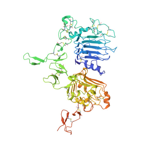
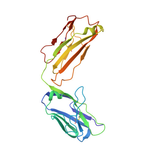
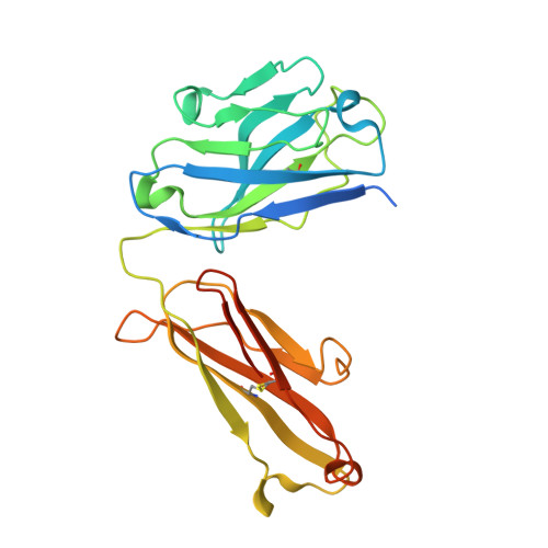
Unbiased Combinatorial Screening Identifies a Bispecific IgG1 that Potently Inhibits HER3 Signaling via HER2-Guided Ligand Blockade.

Geuijen, C.A.W.De Nardis, C.Maussang, D.Rovers, E.Gallenne, T.Hendriks, L.J.A.Visser, T.Nijhuis, R.Logtenberg, T.de Kruif, J.Gros, P.Throsby, M.

(2018) Cancer Cell 33: 922-936.e10

  • DOI: https://doi.org/10.1016/j.ccell.2018.04.003
  • Primary Citation Related Structures: 
    5O4G, 5O4O, 5O7P

  • PubMed Abstract: 

    HER2-driven cancers require phosphatidylinositide-3 kinase (PI3K)/Akt signaling through HER3 to promote tumor growth and survival. The therapeutic benefit of HER2-targeting agents, which depend on PI3K/Akt inhibition, can be overcome by hyperactivation of the heregulin (HRG)/HER3 pathway. Here we describe an unbiased phenotypic combinatorial screening approach to identify a bispecific immunoglobulin G1 (IgG1) antibody against HER2 and HER3. In tumor models resistant to HER2-targeting agents, the bispecific IgG1 potently inhibits the HRG/HER3 pathway and downstream PI3K/Akt signaling via a "dock & block" mechanism. This bispecific IgG1 is a potentially effective therapy for breast cancer and other tumors with hyperactivated HRG/HER3 signaling.


  • Organizational Affiliation
    • Merus NV, 3584 Utrecht, the Netherlands.

Macromolecule Content 

  • Total Structure Weight: 118.2 kDa 
  • Atom Count: 7,472 
  • Modeled Residue Count: 960 
  • Deposited Residue Count: 1,067 
  • Unique protein chains: 3

Macromolecules

Find similar proteins by:|  3D Structure
Entity ID: 1
MoleculeChains  Sequence LengthOrganismDetailsImage
Receptor tyrosine-protein kinase erbB-2A [auth C]606Homo sapiensMutation(s): 0 
Gene Names: ERBB2HER2MLN19NEUNGL
EC: 2.7.10.1
UniProt & NIH Common Fund Data Resources
Find proteins for P04626 (Homo sapiens)
Explore P04626 
Go to UniProtKB:  P04626
PHAROS:  P04626
GTEx:  ENSG00000141736 
Entity Groups
Sequence Clusters30% Identity50% Identity70% Identity90% Identity95% Identity100% Identity
UniProt GroupP04626
Glycosylation
Glycosylation Sites: 3Go to GlyGen: P04626-1
Sequence Annotations
Expand
Reference Sequence
Find similar proteins by:|  3D Structure
Entity ID: 2
MoleculeChains  Sequence LengthOrganismDetailsImage
MF3958 FAB light chainB [auth A]214Homo sapiensMutation(s): 0 
Find similar proteins by:|  3D Structure
Entity ID: 3
MoleculeChains  Sequence LengthOrganismDetailsImage
MF3958 FAB heavy chainC [auth B]247Homo sapiensMutation(s): 0 

Oligosaccharides

Help  
Entity ID: 4
MoleculeChains Length2D Diagram GlycosylationD Interactions
alpha-D-mannopyranose-(1-3)-alpha-D-mannopyranose-(1-6)-[alpha-D-mannopyranose-(1-3)]beta-D-mannopyranose-(1-4)-2-acetamido-2-deoxy-beta-D-glucopyranose-(1-4)-2-acetamido-2-deoxy-beta-D-glucopyranose
D
6N-Glycosylation
Glycosylation Resources
GlyTouCan: G09724ZC
GlyCosmos: G09724ZC
GlyGen: G09724ZC
Entity ID: 5
MoleculeChains Length2D Diagram GlycosylationD Interactions
2-acetamido-2-deoxy-beta-D-glucopyranose-(1-4)-2-acetamido-2-deoxy-beta-D-glucopyranose
E
2N-Glycosylation
Glycosylation Resources
GlyTouCan: G42666HT
GlyCosmos: G42666HT
GlyGen: G42666HT

Small Molecules

Ligands 1 Unique
IDChains Name / Formula / InChI Key2D Diagram3D Interactions
NAG

Query on NAG


Download:Ideal Coordinates CCD File
F [auth C]2-acetamido-2-deoxy-beta-D-glucopyranose
C8 H15 N O6
OVRNDRQMDRJTHS-FMDGEEDCSA-N

Experimental Data & Validation

Experimental Data

  • Method: X-RAY DIFFRACTION
  • Resolution: 3.00 Å
  • R-Value Free:  0.271 (Depositor), 0.274 (DCC) 
  • R-Value Work:  0.229 (Depositor), 0.234 (DCC) 
  • R-Value Observed: 0.231 (Depositor) 
Space Group: P 1 21 1
Unit Cell:
Length ( Å )Angle ( ˚ )
a = 62.79α = 90
b = 113.5β = 101.92
c = 113.49γ = 90
Software Package:
Software NamePurpose
PHENIXrefinement
iMOSFLMdata reduction
Aimlessdata scaling
PHASERphasing

Structure Validation

View Full Validation Report


Ligand Structure Quality Assessment 


Entry History 

Deposition Data

Revision History  (Full details and data files)

  • Version 1.0: 2018-05-16
    Type: Initial release
  • Version 1.1: 2018-05-23
    Changes: Data collection, Database references
  • Version 1.2: 2019-02-20
    Changes: Advisory, Data collection, Derived calculations
  • Version 2.0: 2020-07-29
    Type: Remediation
    Reason: Carbohydrate remediation
    Changes: Atomic model, Data collection, Derived calculations, Structure summary
  • Version 2.1: 2024-01-17
    Changes: Data collection, Database references, Derived calculations, Refinement description, Structure summary
  • Version 2.2: 2024-10-16
    Changes: Structure summary