5O2Z | pdb_00005o2z

Domain swap dimer of the G167R variant of gelsolin second domain


Experimental Data Snapshot

  • Method: X-RAY DIFFRACTION
  • Resolution: 1.70 Å
  • R-Value Free: 
    0.242 (Depositor), 0.242 (DCC) 
  • R-Value Work: 
    0.199 (Depositor), 0.200 (DCC) 
  • R-Value Observed: 
    0.201 (Depositor) 

Starting Model: experimental
View more details

wwPDB Validation 3D Report Full Report

Validation slider image for 5O2Z

This is version 1.4 of the entry. See complete history

Literature

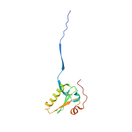
Gelsolin pathogenic Gly167Arg mutation promotes domain-swap dimerization of the protein.

Boni, F.Milani, M.Barbiroli, A.Diomede, L.Mastrangelo, E.de Rosa, M.

(2018) Hum Mol Genet 27: 53-65

  • DOI: https://doi.org/10.1093/hmg/ddx383
  • Primary Citation Related Structures: 
    5O2Z

  • PubMed Abstract: 

    AGel amyloidosis is a genetic degenerative disease characterized by the deposition of insoluble gelsolin protein aggregates in different tissues. Until recently, this disease was associated with two mutations of a single residue (Asp187 to Asn/Tyr) in the second domain of the protein. The general opinion is that pathogenic variants are not per se amyloidogenic but rather that the mutations trigger an aberrant proteolytic cascade, which results in the production of aggregation prone fragments. Here, we report the crystal structure of the second domain of gelsolin carrying the recently identified Gly167Arg mutation. This mutant dimerizes through a three-dimensional domain swapping mechanism, forming a tight but flexible assembly, which retains the structural topology of the monomer. To date, such dramatic conformational changes of this type have not been observed. Structural and biophysical characterizations reveal that the Gly167Arg mutation alone is responsible for the monomer to dimer transition and that, even in the context of the full-length protein, the pathogenic variant is prone to form dimers. These data suggest that, in addition to the well-known proteolytic-dependent mechanism, an alternative oligomerization pathway may participate in gelsolin misfolding and aggregation. We propose to integrate this alternative pathway into the current model of the disease that may also be relevant for other types of AGel amyloidosis, and other related diseases with similar underlying pathological mechanisms.


  • Organizational Affiliation
    • CNR Istituto di Biofisica, c/o Dipartimento di Bioscienze, Università degli Studi di Milano, 20133 Milan, Italy.

Macromolecule Content 

  • Total Structure Weight: 27.25 kDa 
  • Atom Count: 2,079 
  • Modeled Residue Count: 213 
  • Deposited Residue Count: 238 
  • Unique protein chains: 1

Macromolecules

Find similar proteins by:|  3D Structure
Entity ID: 1
MoleculeChains  Sequence LengthOrganismDetailsImage
GelsolinA [auth B],
B [auth A]
119Homo sapiensMutation(s): 1 
Gene Names: GSN
UniProt & NIH Common Fund Data Resources
Find proteins for P06396 (Homo sapiens)
Explore P06396 
Go to UniProtKB:  P06396
PHAROS:  P06396
GTEx:  ENSG00000148180 
Entity Groups
Sequence Clusters30% Identity50% Identity70% Identity90% Identity95% Identity100% Identity
UniProt GroupP06396
Sequence Annotations
Expand
Reference Sequence

Small Molecules

Ligands 5 Unique
IDChains Name / Formula / InChI Key2D Diagram3D Interactions
FLC

Query on FLC


Download:Ideal Coordinates CCD File
D [auth B],
J [auth A]
CITRATE ANION
C6 H5 O7
KRKNYBCHXYNGOX-UHFFFAOYSA-K
GOL

Query on GOL


Download:Ideal Coordinates CCD File
K [auth A]GLYCEROL
C3 H8 O3
PEDCQBHIVMGVHV-UHFFFAOYSA-N
ACT

Query on ACT


Download:Ideal Coordinates CCD File
E [auth B],
F [auth B]
ACETATE ION
C2 H3 O2
QTBSBXVTEAMEQO-UHFFFAOYSA-M
CA

Query on CA


Download:Ideal Coordinates CCD File
C [auth B],
I [auth A]
CALCIUM ION
Ca
BHPQYMZQTOCNFJ-UHFFFAOYSA-N
NA

Query on NA


Download:Ideal Coordinates CCD File
G [auth B],
H [auth B]
SODIUM ION
Na
FKNQFGJONOIPTF-UHFFFAOYSA-N

Experimental Data & Validation

Experimental Data

  • Method: X-RAY DIFFRACTION
  • Resolution: 1.70 Å
  • R-Value Free:  0.242 (Depositor), 0.242 (DCC) 
  • R-Value Work:  0.199 (Depositor), 0.200 (DCC) 
  • R-Value Observed: 0.201 (Depositor) 
Space Group: C 1 2 1
Unit Cell:
Length ( Å )Angle ( ˚ )
a = 106.29α = 90
b = 44.42β = 110.13
c = 58.01γ = 90
Software Package:
Software NamePurpose
XDSdata reduction
XDSdata scaling
PHASERphasing
PHENIXrefinement

Structure Validation

View Full Validation Report


Entry History 

& Funding Information

Deposition Data


Funding OrganizationLocationGrant Number
Fondazione TelethonItalyGEP15070

Revision History  (Full details and data files)

  • Version 1.0: 2017-11-08
    Type: Initial release
  • Version 1.1: 2018-01-03
    Changes: Database references
  • Version 1.2: 2019-08-14
    Changes: Data collection
  • Version 1.3: 2024-01-17
    Changes: Data collection, Database references, Derived calculations, Refinement description
  • Version 1.4: 2024-11-06
    Changes: Structure summary