5O1K | pdb_00005o1k

Crystal structure of murine neuroglobin mutant V140W under 20 bar xenon pressure


Experimental Data Snapshot

  • Method: X-RAY DIFFRACTION
  • Resolution: 2.05 Å
  • R-Value Free: 
    0.202 (Depositor), 0.201 (DCC) 
  • R-Value Work: 
    0.152 (Depositor), 0.164 (DCC) 
  • R-Value Observed: 
    0.155 (Depositor) 

Starting Model: experimental
View more details

wwPDB Validation 3D Report Full Report

Validation slider image for 5O1K

Ligand Structure Quality Assessment 


This is version 1.3 of the entry. See complete history

Literature

Mapping Hydrophobic Tunnels and Cavities in Neuroglobin with Noble Gas under Pressure.

Colloc'h, N.Carpentier, P.Montemiglio, L.C.Vallone, B.Prange, T.

(2017) Biophys J 113: 2199-2206

  • DOI: https://doi.org/10.1016/j.bpj.2017.10.014
  • Primary Citation Related Structures: 
    5NVI, 5NW6, 5O17, 5O18, 5O1K, 5O27, 6EYE

  • PubMed Abstract: 

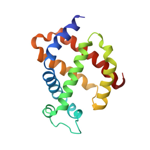
    Internal cavities are crucial for conformational flexibility of proteins and can be mapped through noble gas diffusion and docking. Here we investigate the hydrophobic cavities and tunnel network in neuroglobin (Ngb), a hexacoordinated heme protein likely to be involved in neuroprotection, using crystallography under noble gas pressure, mostly at room temperature. In murine Ngb, a large internal cavity is involved in the heme sliding mechanism to achieve binding of gaseous ligands through coordination to the heme iron. In this study, we report that noble gases are hosted by two major sites within the internal cavity. We propose that these cavities could store oxygen and allow its relay in the heme proximity, which could correspond to NO location in the nitrite-reductase function of Ngb. Thanks to a recently designed pressurization cell using krypton at high pressure, a new gas binding site has been characterized that reveals an alternate pathway for gaseous ligands. A new gas binding site on the proximal side of the heme has also been characterized, using xenon pressure on a Ngb mutant (V140W) that binds CO with a similar rate and affinity to the wild-type, despite a reshaping of the internal cavity. Moreover, this study, to our knowledge, provides new insights into the determinants of the heme sliding mechanism, suggesting that the shift at the beginning of helix G precedes and drives this process.


  • Organizational Affiliation
    • ISTCT CNRS UNICAEN CEA Normandie University, CERVOxy Team, Centre Cyceron, Caen, France. Electronic address: colloch@cyceron.fr.

Macromolecule Content 

  • Total Structure Weight: 18.31 kDa 
  • Atom Count: 1,310 
  • Modeled Residue Count: 148 
  • Deposited Residue Count: 148 
  • Unique protein chains: 1

Macromolecules

Find similar proteins by:|  3D Structure
Entity ID: 1
MoleculeChains  Sequence LengthOrganismDetailsImage
Neuroglobin148Mus musculusMutation(s): 3 
Gene Names: Ngb
EC: 1.7
UniProt
Find proteins for Q9ER97 (Mus musculus)
Explore Q9ER97 
Go to UniProtKB:  Q9ER97
Entity Groups
Sequence Clusters30% Identity50% Identity70% Identity90% Identity95% Identity100% Identity
UniProt GroupQ9ER97
Sequence Annotations
Expand
Reference Sequence

Small Molecules

Ligands 4 Unique
IDChains Name / Formula / InChI Key2D Diagram3D Interactions
HEM

Query on HEM


Download:Ideal Coordinates CCD File
B [auth A]PROTOPORPHYRIN IX CONTAINING FE
C34 H32 Fe N4 O4
KABFMIBPWCXCRK-RGGAHWMASA-L
XE

Query on XE


Download:Ideal Coordinates CCD File
D [auth A]
E [auth A]
F [auth A]
G [auth A]
H [auth A]
D [auth A],
E [auth A],
F [auth A],
G [auth A],
H [auth A],
I [auth A]
XENON
Xe
FHNFHKCVQCLJFQ-UHFFFAOYSA-N
SO4

Query on SO4


Download:Ideal Coordinates CCD File
C [auth A]SULFATE ION
O4 S
QAOWNCQODCNURD-UHFFFAOYSA-L
DIO

Query on DIO


Download:Ideal Coordinates CCD File
J [auth A]1,4-DIETHYLENE DIOXIDE
C4 H8 O2
RYHBNJHYFVUHQT-UHFFFAOYSA-N

Experimental Data & Validation

Experimental Data

  • Method: X-RAY DIFFRACTION
  • Resolution: 2.05 Å
  • R-Value Free:  0.202 (Depositor), 0.201 (DCC) 
  • R-Value Work:  0.152 (Depositor), 0.164 (DCC) 
  • R-Value Observed: 0.155 (Depositor) 
Space Group: H 3 2
Unit Cell:
Length ( Å )Angle ( ˚ )
a = 89.257α = 90
b = 89.257β = 90
c = 115.416γ = 120
Software Package:
Software NamePurpose
SCALAdata scaling
REFMACrefinement
PDB_EXTRACTdata extraction
XDSdata reduction
REFMACphasing

Structure Validation

View Full Validation Report


Ligand Structure Quality Assessment 


Entry History 

Deposition Data

Revision History  (Full details and data files)

  • Version 1.0: 2017-11-15
    Type: Initial release
  • Version 1.1: 2017-11-29
    Changes: Database references
  • Version 1.2: 2018-02-21
    Changes: Structure summary
  • Version 1.3: 2024-01-17
    Changes: Data collection, Database references, Refinement description